

Dual-energy computed tomography derived pulmonary blood volume: association with pulmonary blood flow
- PMID: 41126267
- PMCID: PMC12548200
- DOI: 10.1186/s12931-025-03374-8
Dual-energy computed tomography derived pulmonary blood volume: association with pulmonary blood flow
Abstract
Background: Distribution of ventilation and pulmonary perfusion are the major determinants of pulmonary gas exchange. To study and compare strategies of mechanical ventilation in respiratory research accurate and high-resolution methods are needed to derive distribution of ventilation and perfusion with minimal additional intervention or radiation allowing repeated measurements. Dual-energy computed tomography (DECT) is an imaging technique allowing for the derivation of regional pulmonary perfused blood volume, as a surrogate for pulmonary perfusion (PPDECT). Here accuracy of PPDECT is evaluated in comparison to pulmonary blood flow measured with fluorescence-labeled microspheres (PPFLM). Its feasibility of repeated measurements is evaluated.
Methods: Agreement between PPFLM and PPDECT was assessed by regression as well as Bland-Altman analysis in three anesthetized pigs using DECT and fluorescence labelled microspheres, respectively. Measurements were performed in two-lung and, after right sided thoracotomy, at one-lung ventilation with inhaled nitric oxide. PPFLM and PPDECT were assessed in three different regions of interest (ROI): the right (non-ventilated) and left (ventilated) upper and lower lung, yielding a total of 45 paired measurements over four hours. Persistent iodine accumulation was assessed by additional DECT scans before each contrast administration.
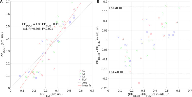
Results: Regression analysis revealed a good overall association (R2 = 0.81) between PPFLM and PPDECT, with PPDECT substantially overestimating PPFLM up to 30%, with limits of agreement of -18 and 18%, Low PPFLM was underestimated, while high PPFLM was overestimated by PPDECT, indicating a higher sensitivity of the later. Changes of PPDECT and PPFLM had a concordance of 69.4% for all measurements. Agreement and concordance were highest in ventilated and lowest in non-ventilated ROIs. No persistent iodine enhancement was detected in the lung parenchyma after repetitive measurements per hour.
Conclusions: Dual-energy CT based measurement of pulmonary perfusion shows promising results indicating its feasibility in translational research on strategies of mechanical ventilation.
Keywords: Dual-energy CT; Fluorescence labelled microspheres; Regional pulmonary perfusion; Shunt blood flow.
© 2025. The Author(s).
Conflict of interest statement
Declarations. Ethics approval and consent to participate: The protocol was approved by the Institutional Animal Care and Welfare Committee and the Government of Saxony, Germany (file: 25–5131/496/33) and the Animal Research: Reporting of In Vivo Experiments (ARRIVE) guidelines were followed. Consent for publication: Not applicable. Competing interests: The authors of this manuscript declare relationships with the following companies: MGA: Dräger Medical, Ambu, GE Healthcare, and ZOLL. The other authors of this manuscript declare no relationships with any companies, whose products or services may be related to the subject matter of the article. The other authors of this manuscript declare no relationships with any companies, whose products or services may be related to the subject matter of the article.
Figures


References
-
- Almeida IP, Schyns LEJR, Öllers MC, van Elmpt W, Parodi K, Landry G, et al. Dual-energy CT quantitative imaging: a comparison study between twin-beam and dual-source CT scanners. Med Phys. 2017;44(1):171–9. - PubMed
MeSH terms
LinkOut - more resources
Full Text Sources
Medical
Miscellaneous
