. 2025 Oct 16:18:14325-14341.
doi: 10.2147/JIR.S544625. eCollection 2025.

Chronic Sleep Fragmentation Differentially Affects Alzheimer's Disease Pathology in Male and Female APPSAA Knock-in Mice

Affiliations

Chronic Sleep Fragmentation Differentially Affects Alzheimer's Disease Pathology in Male and Female APPSAA Knock-in Mice

Margaret R Hawkins et al. J Inflamm Res. .

Abstract

Introduction: Sleep fragmentation often precedes Alzheimer's disease (AD) diagnosis and represents a potential modifiable risk factor, especially among women who have higher prevalence of both sleep disorders and AD.

Methods: This study investigated how chronic sleep fragmentation affects neuroinflammation and amyloid-beta (Aβ) accumulation in male and female APPSAA knock-in mice, a physiologically relevant AD model expressing APP at normal levels. APPSAA mice of both sexes (N=8/sex/strain, 8 months old) underwent either 5 weeks of chronic sleep fragmentation administered during the light phase using an automated sweeper system or undisturbed sleep. Sleep-wake patterns and circadian rhythms were monitored using piezoelectric sensors. Following intervention, we assessed neuroinflammatory markers via immunohistochemistry and multiplex cytokine analysis, Aβ levels in different solubility fractions, and Aβ plaque characteristics through digital pathology.

Results: Sleep fragmentation effectively disrupted sleep patterns in both sexes, reducing light-phase sleep and increasing intradaily variability. Sleep fragmentation increased GFAP immunoreactivity in both sexes, with larger effects in females than males. Surprisingly, sleep fragmentation decreased expression of the microglial activation markers MHCII and Dectin-1 in males. Pro-inflammatory cytokines (IL-1β, CCL2, CXCL2) were significantly elevated following sleep fragmentation, with distinct regional and sex-specific patterns. In females, sleep fragmentation increased PBS-soluble and formic acid-soluble Aβ in the neocortex and medium-sized plaque density in the hippocampus, while males showed decreased detergent-soluble Aβ in the neocortex following sleep fragmentation.

Discussion: Chronic sleep fragmentation exacerbates AD-related pathology in APPSAA mice in a sex-dependent manner, with females showing greater vulnerability to Aβ accumulation and astrocyte reactivity following sleep disruption. These findings suggest that environmental sleep disruptions may contribute to the higher prevalence of AD in women and highlight the importance of addressing sleep fragmentation as a modifiable risk factor for AD.

Keywords: amyloidosis; astrocyte reactivity; circadian rhythm; environmental stressors; neuroinflammation; sexual dimorphism.


Conflict of interest statement

BFO is a co-owner of Signal Solutions, LLC, which manufactures the piezoelectric sleep monitoring equipment used in this study. He also reports NIH grants to Signal Solutions, typically with 50% subawarded to UK, but not directly related to this manuscript (but potentially synergistic). On his current STTR award, he is the PI through his UK faculty position, even though the award is to Signal Solutions. He also reports multiple conflict of interest management plans, and a UK COI committee, to help him manage these potential conflicts of interest. He receives a portion of his yearly income from Signal Solutions, that is variable, but averages roughly 25% of his yearly salary. He attends most scientific meetings paid by Signal Solutions, even when presenting UK related research, mainly because it is easier; non-financial support includes admin support from his company, to free up his time for both UK and the company; reports patent “Stimulation System Based on Mechanical Vibration for Modification and Characterization of Sleep and Behavior in Rodents”, not directly relevant to the manuscript. MJD reports honoraria for reviews of grants related to research on Alzheimer’s disease from State of Florida Department of Health. All other authors report no conflicts of interest related to this work.

Figures

Figure 1
Figure 1
Sleep fragmentation disrupts sleep-wake patterns and circadian rhythms in APPSAA mice. (A) Twenty-four-hour sleep profiles showing percentage of time spent asleep throughout the day (Zeitgeber time) in female (top) and male (bottom) mice at baseline, week 1 of fragmentation, week 4 rest period, and week 5 of fragmentation. Yellow lines represent undisturbed sleep (US) controls; purple lines represent sleep fragmentation (SF) groups. Shaded areas indicate the dark phase (active period). Data shown as mean with shaded regions representing SD. Vertical dotted lines indicate light-dark transitions. (B) Percentage of time spent asleep during the light phase (left), dark phase (middle), and total 24-hour period (right) at weeks 1 and 5. Sleep fragmentation significantly decreased sleep during the light phase while increasing sleep during the dark phase, particularly in females at week 1. (C) Intradaily variability (IV), a measure of rhythm fragmentation, showing significant increases with sleep fragmentation in both sexes at weeks 1 and 5. Higher values indicate more disrupted and fragmented daily rhythms, a characteristic associated with neurodegenerative conditions. (D) Mean 24-hour activity level (MESOR) showing increased overall activity with sleep fragmentation, with a significant sex-by-treatment interaction at week 5. (E) Amplitude of the activity rhythm (peak-to-trough difference) showing pronounced reduction with sleep fragmentation, indicating flattening of the circadian rhythm. The effect was more persistent in males at week 5. Data in (A) are presented as mean ± SD. Data in (B-E) are presented as mean ± SEM with individual data points (n = 8 per group). P-values from post-hoc analyses between US and SF conditions are indicated.
Figure 2
Figure 2
Sleep fragmentation alters Aβ levels and plaque characteristics in APPSAA mice. (A) ELISA measurements of Aβ in PBS-soluble (left), detergent-soluble (middle), and formic acid-soluble (right) fractions from neocortex and hippocampus. Sleep fragmentation increased PBS-soluble and formic acid-soluble Aβ in female neocortex while decreasing detergent-soluble Aβ in the hippocampus of both sexes. (B) Representative images of 6E10 immunostaining showing Aβ plaques in male and female APPSAA mice with undisturbed sleep (US) or sleep fragmentation (SF). Scale bars: 25 μm. (C) Quantification of total Aβ plaque burden (% area) measured by digital pathology using the HALO area fraction algorithm. (D) Plaque density (number/mm²) by size category: small (10–99 μm²), medium (100–399 μm²), and large (≥400 μm²) plaques, quantified using the HALO object colocalization algorithm with size exclusions. Sleep fragmentation significantly increased medium-sized plaque density in female hippocampus. Data presented as mean ± SEM with individual data points (n = 8 per group).
Figure 3
Figure 3
Sleep fragmentation increases neuroinflammatory cytokines and chemokines in APPSAA mice. (A) Heatmap displaying z-score normalized levels of cytokines and chemokines in the neocortex and hippocampus of female and male APPSAA mice with undisturbed sleep (US) or sleep fragmentation (SF). Yellow-Orange indicates higher expression while purple-black indicates lower expression relative to the mean across all groups. Bold and asterisk in the cytokine name indicate the main effect of sleep fragmentation (*p<0.05, **p<0.01). In the heatmap boxes, asterisks indicate significant differences (p < 0.05) between US and SF conditions within each sex and brain region. (B) Cumulative inflammatory z-score representing the overall cytokine/chemokine burden in neocortex and hippocampus. Sleep fragmentation significantly increased the inflammatory index in both female and male neocortex. (CE) Absolute concentrations (pg/mg total protein) of representative inflammatory mediators: IL-1β (C), CCL2 (D), and CXCL2 (E). Data presented as mean ± SEM with individual data points (n = 8 per group).
Figure 4
Figure 4
Sleep fragmentation increases astrocyte reactivity in both male and female APPSAA mice. (A) Representative images of GFAP immunostaining showing distribution across brain sections, with higher magnification images of the neocortex and hippocampus from male and female APPSAA knock-in mice with undisturbed sleep (US) or sleep fragmentation (SF). Scale bars: 500 μm (top row), 50 μm (middle and bottom rows). (B) Example of HALO digital pathology quantification showing area fraction markup of GFAP-positive staining (yellow). Scale bars: 10 μm. (C) Quantification of GFAP-positive area (%) in the neocortex and hippocampus of female and male APPSAA mice with US or SF (n = 8 per group). Quantification was performed at 300 μm intervals throughout each brain region using HALO digital pathology software. Data are presented as mean ± SEM with individual data points shown. Sleep fragmentation increased GFAP immunoreactivity in the neocortex of females (52.2% increase, Cohen’s d = 2.47) and males (36.2% increase, Cohen’s d = 1.97), and in the hippocampus of females (28.1% increase, Cohen’s d = 2.13) and males (20.9% increase, Cohen’s d = 1.44). P-values from planned post-hoc analyses between US and SF conditions are indicated.
Figure 5
Figure 5
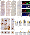
Sleep fragmentation selectively reduces MHCII and Dectin-1 expression in male APPSAA mice. (A) Representative low-magnification images show the pattern of IBA1, CD45, MHCII, and Dectin-1 staining across the neocortex, hippocampus, and deeper structures such as the thalamus, with the most robust staining observed in the neocortex and hippocampus. (B) Widefield epifluorescence images show that MHCII colocalizes with IBA1⁺ microglia but not GFAP⁺ astrocytes. (C) Dectin-1 also colocalizes with IBA1⁺ microglia but not GFAP⁺ astrocytes. (D) Representative images illustrate distinct microglial/macrophage phenotypes in APPSAA mice by treatment group. IBA1 reveals plaque-associated microglia with hypertrophic morphology; CD45 identifies potential infiltrating leukocytes, particularly in the female hippocampus; MHCII labels antigen-presenting cells, predominantly in the male hippocampus under undisturbed sleep; and Dectin-1 marks phagocytic cells associated with damage-responsive states. Arrow highlights a Dectin-1⁺ cell that appears to be engulfing another cell. Halo digital pathological quantification of IBA1 (E), CD45 (F), MHCII (G), and Dectin-1 (H) in neocortex and hippocampus. Scale bars: A = 500 μm; B–D = 25 μm.

References

    1. Dufort-Gervais J, Mongrain V, Brouillette J. Bidirectional relationships between sleep and amyloid-beta in the hippocampus. Neurobiol Learn Mem. 2019;160:108–117. doi: 10.1016/j.nlm.2018.06.009 - DOI - PubMed
    1. Yes J, Lucey BP, Holtzman DM. Sleep and Alzheimer disease pathology-a bidirectional relationship. Nat Rev Neurol. 2014;10(2):115–119. doi: 10.1038/nrneurol.2013.269 - DOI - PMC - PubMed
    1. Sterniczuk R, Dyck RH, Laferla FM, Antle MC. Characterization of the 3xTg-AD mouse model of Alzheimer’s disease: part 1. Circadian changes. Brain Res. 2010;1348:139–148. doi: 10.1016/j.brainres.2010.05.013 - DOI - PubMed
    1. Ambree O, Touma C, Gortz N, et al. Activity changes and marked stereotypic behavior precede Abeta pathology in TgCRND8 Alzheimer mice. Neurobiol Aging. 2006;27(7):955–964. doi: 10.1016/j.neurobiolaging.2005.05.009 - DOI - PubMed
    1. Kang JE, Lim MM, Bateman RJ, et al. Amyloid-beta dynamics are regulated by orexin and the sleep-wake cycle. Science. 2009;326(5955):1005–1007. doi: 10.1126/science.1180962 - DOI - PMC - PubMed

LinkOut - more resources