. 2025 Oct 23;16(1):9374.
doi: 10.1038/s41467-025-64426-7.

Targeted degradation of endogenous YAP by nanobody bioPROTAC inhibits tumor progression

Affiliations

Targeted degradation of endogenous YAP by nanobody bioPROTAC inhibits tumor progression

Runhua Zhou et al. Nat Commun. .

Abstract

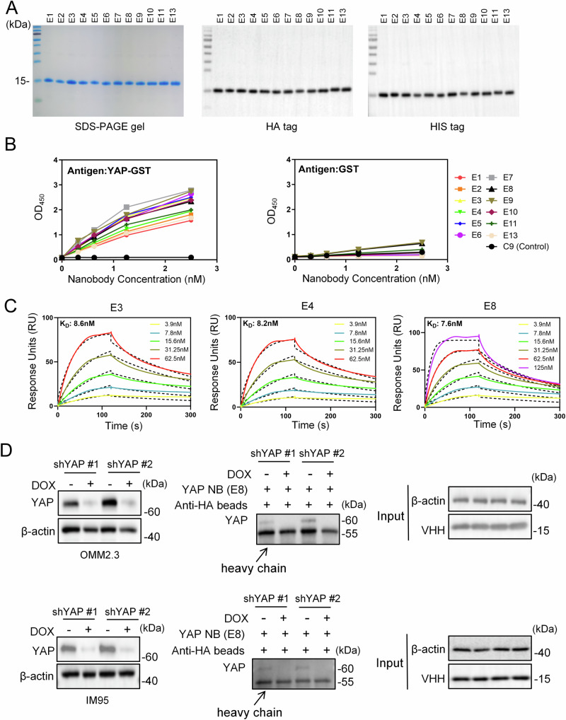
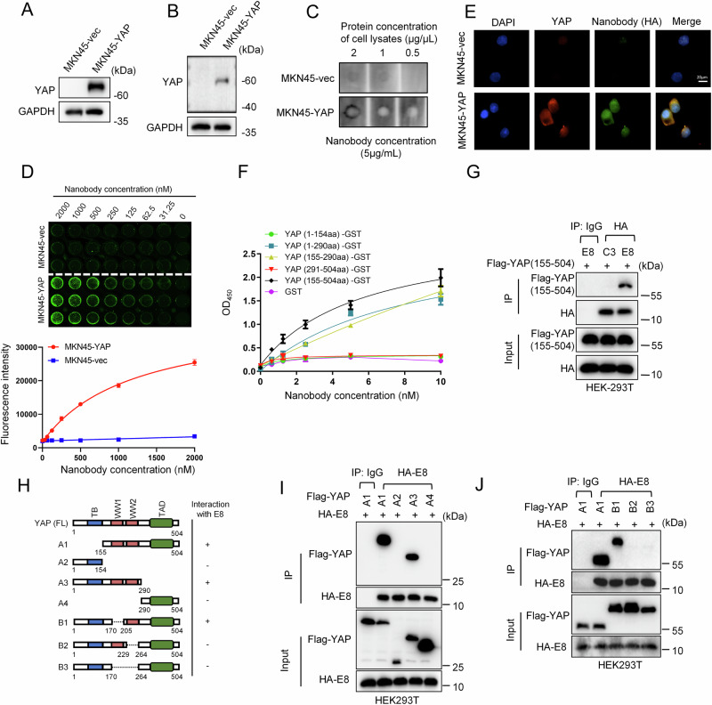
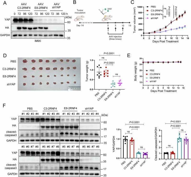
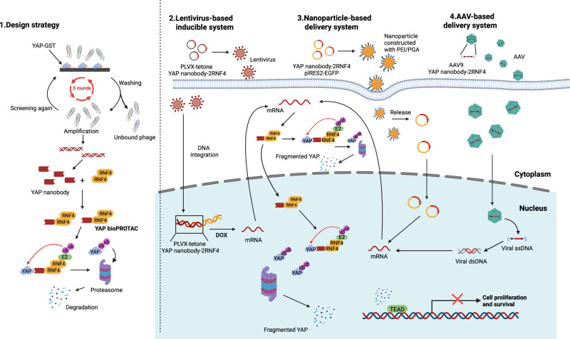
Yes-associated protein (YAP), a key effector of the Hippo pathway, regulates gene expression and promotes tumorigenesis. YAP is conventionally considered "undruggable", however, targeted protein degradation offers a promising approach to address the challenges associated with targeting this oncogenic protein. In this study, through naïve nanobody phage library screening, we identify multiple nanobodies against human YAP with high affinity and specificity. The YAP nanobody is then fused to the RING domain of RNF4, creating a bio-Proteolysis-Targeting Chimera (bioPROTAC) molecule capable of selectively targeting endogenous YAP for ubiquitin-mediated degradation. Notably, the constructed YAP bioPROTAC demonstrates significant YAP degradation and anticancer efficacy in various YAP-dependent cancers both in vitro and in vivo. Nanoparticles and adeno-associated virus (AAV) can effectively deliver the encoding gene of YAP bioPROTAC, achieving YAP degradation in tumors. Collectively, our study provides a proof-of-concept that the YAP nanobody-bioPROTAC approach can effectively degrade endogenous YAP via the ubiquitin-proteasome system, highlighting a feasible strategy for "undruggable" YAP-dependent cancers.


Conflict of interest statement

Competing interests: The authors declare no competing interests.

Figures

Fig. 1
Fig. 1. Screening of nanobodies targeting YAP proteins using a naïve phage nanobody library.
A SDS-PAGE analysis of recombinant YAP-GST protein and the control GST protein. Data are representative of two independent experiments. B Phage titer measurement of eluates from each round of panning against YAP-GST, with negative selection against GST on the 3rd round of panning. C, D Phage ELISA experiments with 192 randomly selected clones, showing 72 clones with YAP-GST/GST OD450 value ratios >3, and 120 clones with ratios <3. E Two highly enriched CDR3 regions from 12 unique nanobody sequences.
Fig. 2
Fig. 2. Purification and affinity determination of YAP-specific nanobodies.
A Purification and Western blot analysis of 12 nanobodies identified from phage ELISA. Data are representative of two independent experiments. B ELISA analysis evaluating the binding ability of 12 nanobodies and a negative control nanobody (C9) to YAP-GST and GST. Data from one representative experiment out of two performed is depicted. C Determination of nanobody E3/E4/E8 binding affinity to YAP-GST protein using Surface Plasmon Resonance (SPR). Solid lines represent the experimental sensorgrams and dashed lines indicate the fitted curves. The equilibrium dissociation constants (KD) were measured as 8.6 nM (E3), 8.2 nM (E4), and 7.6 nM (E8), respectively. D Validation of the specific interaction between nanobody E8 and endogenous YAP protein through pull-down assays. YAP was downregulated in OMM2.3 and IM95 cells using doxycycline (DOX)-inducible (Tet-on) shRNA targeting YAP. Cells were treated with or without DOX (500 ng/ml) for 48 h. Western blot analysis was performed to determine YAP protein pulled down by HA-tagged beads from cell lysates containing nanobody E8. The total protein levels were assessed using anti-actin and anti-VHH antibodies (input). Data are representative of two independent experiments.
Fig. 3
Fig. 3. YAP Nanobody-bioPROTAC fusions induce intracellular YAP degradation and suppress tumor cell growth.
Time-course experiments determining the degradation of YAP in uveal melanoma cells (OMM2.3 and 92.1) (A) gastric cancer cells (IM95 and AGS) (B) breast cancer cells (MDA-MB-231) (C) mesothelioma cells (NCI-H2373, NCI-H2052 and MSTO-211H) (D) following inducible expression of C3 or E8 nanobody-2RNF4 upon doxycycline (DOX) treatment (200 ng/ml). Quantification data are presented as mean ± SEM from three independent experiments. Colony formation assays validating the growth inhibitory effects of E8-2RNF4 on OMM2.3, 92.1 (E), IM95, AGS (F), MDA-MB-231 (G), NCI-H2373, NCI-H2052, MSTO-211H (H) cells (n = 3 biological replicates, mean ± SEM, two-sided Student’s t-test). I Cell apoptosis analysis by flow cytometry after staining with Annexin V-FITC and PI. Cells stably expressing inducible E8-2RNF4 or C3-2RNF4 were treated with DOX (200 ng/ml) for 48 h (n = 3 biological replicates, mean ± SEM, two-sided Student’s t-test, “ns” represents no significant difference). Amino acid sequences of E8-2RNF4 and C3-2RNF4 are available in Supplementary Note 1. Source data are provided as a Source Data file.
Fig. 4
Fig. 4. Specific binding of E8 nanobody to YAP.
A Ectopic expression of YAP in MKN45 YAP-null cells was confirmed by western blot. MKN45 cells stably expressing either an empty vector (MKN45-vec) or YAP (MKN45-YAP) were generated by lentiviral infection using pLVX-empty vector or pLVX-YAP constructs. B Western blot analysis of E8 nanobody binding using cell lysates from MKN45-vec or MKN45-YAP. C Dot blot assay examining the binding of E8 to cell lysates from MKN45-vec or MKN45-YAP. D In-Cell ELISA analysis showing the binding of E8 nanobody to MKN45-YAP cells but not MKN45-vec cells (n = 3 biological replicates, mean ± SEM). E Immunofluorescence analysis showing specific recognition of E8 nanobody to YAP protein in MKN45-YAP cells, but not in MKN45-vec cells. Scale bar: 20 μm. F ELISA analysis evaluating the binding ability of E8 to recombinant YAP fragment proteins (n = 2 biological replicates, mean ± SEM). GST served as a negative control. Data are representative of two independent experiments. G Co-immunoprecipiatation (coIP) demonstrating the interaction between Flag-YAP (155-504 aa) and HA-E8. Flag-YAP was cotransfected with HA-C3 (irrelevant control nanobody) or HA-E8 in HEK293T cells. Immunoprecipitated HA-tagged proteins were analyzed for co-precipitation of Flag-YAP by Western blot. H Schematic diagram of YAP deletion constructs (A1/2/3/4 and B1/2/3). TB, TEAD-binding domain; TAD, C-terminal transactivation domain. I The YAP region spanning 155–290 aa is required for its interaction with E8. YAP deletion constructs A1/2/3/4 were cotransfected with HA-E8 into HEK293T cells, and coIP was performed to identify interacting regions. J WW2 domain of YAP mediates its interaction with E8. CoIP experiments, similar to (I), were performed using YAP deletion mutants B1/2/3. Experiments in figures (AC, E, G, I, J) were repeated twice or more. Source data are provided as a Source Data file.
Fig. 5
Fig. 5. YAP bioPROTAC inhibits tumor cell migration in vitro and in vivo.
A Representative images of migrating cells following the inducible expression of E8-2RNF4 with doxycycline (DOX). Quantitative analysis of migrating cells is shown on the right (n = 3, five independent fields for each membrane, mean ± SEM, two-sided Student’s t-test). B, C E8-2RNF4 expression suppresses the dissemination of 92.1 and OMM2.3 cells in zebrafish embryos exposed to DOX. Quantitative analysis of tumor cell foci that migrated outside the eye is shown in (C) (n = 26 in the 92.1 control group; n = 22 in the 92.1 DOX-treated group; n = 26 in the OMM2.3 control group; n = 30 in the OMM2.3 DOX-treated group; mean ± SEM, two-sided Student’s t-test). Source data are provided as a Source Data file.
Fig. 6
Fig. 6. YAP nanobody bioPROTAC arrests tumor growth in vivo.
AC E8-2RNF4 inhibits tumor growth in multiple xenograft models. MDA-MB-231 (C3-2RNF4 or E8-2RNF4), MSTO-211H (E8-2RNF4), and IM95 (E8-2RNF4) cells were subcutaneously implanted into immunodeficient mice to establish multiple xenograft models. Once average tumor size reached ~100 mm3, the animals were randomly grouped and given drinking water with/without doxycycline (DOX, 0.5 mg/ml). n = 7 (MDA-MB-231 and MSTO-211H), n = 8 (IM95). Data are mean ± SEM, two-way ANOVA with Tukey’s test for multiple comparisons, “ns” represents no significant difference. DF E8-2RNF4 prolongs the survival of animals in the xenografted models shown in (AC). Two-sided Mantel–Cox log-rank test with Holm-Sidak correction for multiple comparisons, “ns” represents no significant difference. G Tumor growth curves of the 92.1 (E8-2RNF4) xenograft model. 92.1 (E8-2RNF4) cells were subcutaneously implanted into BALB/c nude mice. Once average tumor size reached ~100 mm3, the animals were randomly grouped and given drinking water with/without DOX (0.5 mg/ml) (n = 8 biological replicates, mean ± SEM, two-way ANOVA). H Images and weights of the isolated tumor tissues from 92.1 tumor-bearing mice after euthanasia (n = 8 biological replicates, mean ± SEM, two-sided Student’s t-test). I Representative immunohistochemical images for HA, YAP and Ki-67 expression in tumors from the 92.1 (E8-2RNF4) xenograft model. Quantification of YAP and Ki-67 expression is shown on the right. The DOX-treated group exhibited a notable increase in HA expression and a concurrent decrease in both YAP and Ki-67 expression (n = 3 biological replicates, mean ± SEM, two-sided Student’s t-test). Source data are provided as a Source Data file.
Fig. 7
Fig. 7. Gene delivery of YAP bioPROTAC encoding plasmid using PEI/PGA-based nanocomplexes inhibits YAP-dependent tumors.
A Western blot analysis of plasmid transfection using PEI or PEI/PGA-based nanocomplexes in MSTO-211H cells. B Growth curves of MSTO-211H tumor xenografts. Mice were treated with various nanocomplexes (E8-2RNF4, C3-2RNF4) or PBS via peritumoral injection. n = 5 (PBS, C3-2RNF4); n = 6 (E8-2RNF4). Data are mean ± SEM, two-way ANOVA followed by Tukey’s test. C Body weight of mice from the groups shown in (B). D Western blot analysis of tumor samples extracted from MSTO-211H-bearing mice after various treatments. E8-2RNF4 nanoparticles significantly down-regulated YAP protein levels and upregulated apoptosis indicator cleaved caspase 3 compared to both the PBS and C3-2RNF4 groups. Quantitative analysis of key indicators is presented on the right. n = 5 (PBS, C3-2RNF4); n = 6 (E8-2RNF4). Data are mean ± SEM, two-sided Student’s t-test. E Western blot analysis of plasmid transfection using PEI or PEI/PGA-based nanocomplexes in IM95 cells. F Growth curves of IM95 tumor xenografts. Mice were treated with nanocomplexes (E8-2RNF4) or PBS via intraperitoneal injection (n = 4 biological replicates, mean ± SEM, two-way ANOVA). G Tumor images and tumor weights in mice from the groups shown in (F) (n = 4 biological replicates, mean ± SEM, two-sided Student’s t-test). Experiments in (A, E) were repeated twice or more. Source data are provided as a Source Data file.
Fig. 8
Fig. 8. AAV9-E8-2RNF4 reduces YAP expression and suppresses tumor growth in vivo.
A IM95 cells were incubated with AAV9-C3-2RNF4, AAV9-E8-2RNF4, or AAV9-YAP shRNA viral particles at a MOI of 105 GC per cell. YAP expression was assessed by western blot at 72, 96, and 120 hr post-transduction. Data from one representative experiment out of two performed is depicted. B Schematic diagram of intratumoral AAV9 treatment. Figure created in BioRender. Jiayun, C. (2025) https://BioRender.com/4b2nb1j. C Growth curves of IM95 tumor xenografts in mice treated with AAV9-C3-2RNF4, AAV9-E8-2RNF4, or AAV9-YAP shRNA via intratumoral injection (n = 8 biological replicates, mean ± SEM, two-way ANOVA followed by Tukey’s test, “ns” represents no significant difference). D Tumor images and tumor weights from the mice shown in (C) (n = 8 biological replicates, mean ± SEM, one-way ANOVA followed by Tukey’s test, “ns” represents no significant difference). E Body weights of mice during the course of treatment as shown in (B). F Western blot analysis of tumor samples extracted from IM95-bearing mice after various treatments. Quantitative analysis of YAP and cleaved caspase 3 levels is presented on the right (n = 8 biological replicates, mean ± SEM, one-way ANOVA followed by Tukey’s test, “ns” represents no significant difference). Source data are provided as a Source Data file.
Fig. 9
Fig. 9. Schematic overview of the design and therapeutic application of a YAP-targeting nanobody-based bioPROTAC.
(1) High-affinity nanobodies against YAP were identified by phage display screening. (2) The selected nanobodies were fused to the RING domain of RNF4 to generate a YAP bioPROTAC, expressed using a lentivirus-based doxycycline-inducible system. (3) PEI/PGA-based nanoparticles were used to encapsulate and deliver the YAP bioPROTAC gene in vivo. (4) An adeno-associated virus (AAV9) system was also employed for efficient gene delivery. The expressed bioPROTAC directs YAP for ubiquitin–proteasome degradation, leading to suppression of YAP/TEAD transcriptional activity and tumor growth. Figure created in BioRender. Jiayun, C. (2025) https://BioRender.com/4b2nb1j.

References

    1. Ma, S., Meng, Z., Chen, R. & Guan, K. L. The Hippo pathway: biology and pathophysiology. Annu. Rev. Biochem.88, 577–604 (2019). - PubMed
    1. Dong, J. et al. Elucidation of a universal size-control mechanism in Drosophila and mammals. Cell130, 1120–1133 (2007). - PMC - PubMed
    1. Zhao, B., Li, L., Tumaneng, K., Wang, C. Y. & Guan, K. L. A coordinated phosphorylation by Lats and CK1 regulates YAP stability through SCF(beta-TRCP). Genes Dev.24, 72–85 (2010). - PMC - PubMed
    1. Zhao, B. et al. Inactivation of YAP oncoprotein by the Hippo pathway is involved in cell contact inhibition and tissue growth control. Genes Dev.21, 2747–2761 (2007). - PMC - PubMed
    1. Franklin, J. M., Wu, Z. & Guan, K. L. Insights into recent findings and clinical application of YAP and TAZ in cancer. Nat. Rev. Cancer23, 512–525 (2023). - PubMed

MeSH terms

LinkOut - more resources