Case Reports
. 2025 Oct 8:12:1683444.
doi: 10.3389/fcvm.2025.1683444. eCollection 2025.

Biopsy-proven acute eosinophilic myocarditis as the initial manifestation of severe primary Sjögren's syndrome: a case report

Affiliations
Case Reports

Biopsy-proven acute eosinophilic myocarditis as the initial manifestation of severe primary Sjögren's syndrome: a case report

Katsuya Hashimoto et al. Front Cardiovasc Med. .

Abstract

Background: Primary Sjögren's syndrome (pSS) is a chronic autoimmune inflammatory disorder primarily affecting the exocrine glands. A subset of patients exhibits extraglandular manifestations, including cardiovascular involvement. Among them, myocarditis is a rare complication, and its pathogenesis remains poorly understood.

Case presentation: An 85-year-old man with a persistent dry mouth was admitted to our hospital with high-grade fever, nausea, fatigue, and urinary disturbance. On day 2, the patient developed multiple cerebral infarctions and bilateral acute otitis media. Fever and inflammatory response without leukocytosis and cardiac imaging findings indicative of active myocarditis, and normal cardiac function suggested acute viral myocarditis, for which supportive treatments were initiated. On day 6, the patient experienced acute heart failure with severely reduced ejection fraction and cardiogenic shock. An endomyocardial biopsy was performed following transient peripheral eosinophilia in serial blood samples, which revealed acute eosinophilic myocarditis (AEM). A thorough diagnostic evaluation for eosinophilia revealed pSS, leading to the final diagnosis of pSS-associated AEM. Systemic high-dose corticosteroid treatment improved the general condition of the patient, except for a left ventricular apical aneurysm. Nevertheless, the patient's post-treatment hospital course was complicated by serious digestive involvement, leading to death from septic shock.

Conclusions: To our knowledge, this is the first case of severe pSS complicated by AEM. This case highlights the importance of early therapeutic intervention for AEM and early comprehensive surveillance of systemic organs for pSS. Furthermore, this case provides new insights into the pathogenesis of pSS-associated myocarditis.

Keywords: acute eosinophilic myocarditis; acute otitis media; digestive involvement; primary Sjögren's syndrome; ulcerative colitis.


Conflict of interest statement

The authors declare that the research was conducted in the absence of any commercial or financial relationships that could be construed as a potential conflict of interest.

Figures
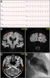
Figure 1
Figure 1
Initial examinations. (A) Initial electrocardiography. (B, C) Coronal views of brain diffusion-weighted magnetic resonance images (DWIs) showing multiple acute cerebral infarctions (arrows). (D) T2-weighted image (T2WI) showing fluid collection as a hyperintense area in bilateral mastoid cavities (arrowheads). (E) Left ventriculogram on day 2 after admission showing normal hyperkinetic contractions with an ejection fraction of 60%.
Figure 2
Figure 2
Enhanced computed tomography (CT) and cardiac magnetic resonance imaging (CMR) (A,C, apical four-chamber view; B,D, long-axis view). CT showing low-attenuation areas of the left ventricular apex (arrowheads). T2-weighted image (T2WI) showing diffuse myocardial edema. Late gadolinium enhancement (LGE) images showing the same region as that of CT (arrowheads). Serial transthoracic echocardiography (TTE). (E,G, apical two-chamber view; and F,H, apical four-chamber view). (E,F) TTE on day 6 after admission showing severe global left ventricular systolic dysfunction with increased left ventricular wall thickness (LVWT) accompanied by severe mitral regurgitation (arrow) in color Doppler mode [LVWT, 14 mm; and ejection fraction (EF), 40%]. (G,H) Follow-up TTE 2 weeks after corticosteroid treatment showing considerable improvement in left ventricular function and mitral regurgitation (LVWT, 12 mm; and EF, 57%). Note the left ventricular apical aneurysmal formation (arrowheads). LA, left atrium; LV, left ventricle; RA, right atrium; RV, right ventricle.
Figure 3
Figure 3
Endomyocardial biopsy. (A) Hematoxylin and eosin staining. (B) Direct fast scarlet staining showing intense eosinophilic infiltration. The inlet shows a magnified view of degranulated eosinophilic granules (arrowhead). (C) Immunostaining for major basic proteins (MBP). (D–F) Immunohistochemistry showing abundant inflammatory infiltrates comprising CD3 + and CD4+ T-lymphocytes. A small number of CD8+ T lymphocytes are also observed. (G, H) A considerable number of CD163 + and CD209+ M2 macrophages are observed. Scale bars: 20 μm.
Figure 4
Figure 4
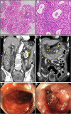
Lip gland histopathology, computed tomography (CT) and colonoscopy findings. (A) Pathology of a minor salivary gland biopsy (20× magnification). (B) Magnified view of the box in (A) at 40×. Hematoxylin and eosin staining showing focal lymphocytic sialadenitis with periductular aggregates of >50 lymphocytes. (C,D) Coronal enhanced CT revealing whole and homogeneous large bowel wall thickening characterized by submucosal edema and mucosal thickening with the disappearance of the haustral folds (arrowheads). Note the intact small bowel. (E,F) Colonoscopy revealed continuous diffuse mucosal inflammation from the rectum to the transverse colon. There are no vascular patterns, multiple ulcers with friability, mucosal desquamation (arrowhead), or contact bleeding.

References

    1. Qin B, Wang J, Yang Z, Yang M, Ma N, Huang F, et al. Epidemiology of primary sjogren’s syndrome: a systematic review and meta-analysis. Ann Rheum Dis. (2015) 74:1983–9. 10.1136/annrheumdis-2014-205375 - DOI - PubMed
    1. Mihai A, Caruntu C, Jurcut C, Blajut FC, Casian M, Opris-Belinski D, et al. The Spectrum of extraglandular manifestations in primary Sjogren’s syndrome. J Pers Med. (2023) 13:961. 10.3390/jpm13060961 - DOI - PMC - PubMed
    1. Atzeni F, Gozza F, Cafaro G, Perricone C, Bartoloni E. Cardiovascular involvement in Sjogren’s syndrome. Front Immunol. (2022) 13:879516. 10.3389/fimmu.2022.879516 - DOI - PMC - PubMed
    1. Shiboski CH, Shiboski SC, Seror R, Criswell LA, Labetoulle M, Lietman TM, et al. 2016 American college of rheumatology/European league against rheumatism classification criteria for primary sjogren’s syndrome: a consensus and data-driven methodology involving three international patient cohorts. Ann Rheum Dis. (2017) 76:9–16. 10.1136/annrheumdis-2016-210571 - DOI - PubMed
    1. Watanabe T, Takahashi Y, Hirabayashi K, Tomaru U, Machida M. Acute fulminant myocarditis in a patient with primary sjogren’s syndrome. Scand J Rheumatol. (2019) 48:164–5. 10.1080/03009742.2018.1514068 - DOI - PubMed

Publication types

LinkOut - more resources