. 2025 Oct 24;16(1):9358.
doi: 10.1038/s41467-025-64540-6.

Development of [18F]ACI-19626 as a first-in-class brain PET tracer for imaging TDP-43 pathology

Affiliations

Development of [18F]ACI-19626 as a first-in-class brain PET tracer for imaging TDP-43 pathology

Efthymia Vokali et al. Nat Commun. .

Abstract

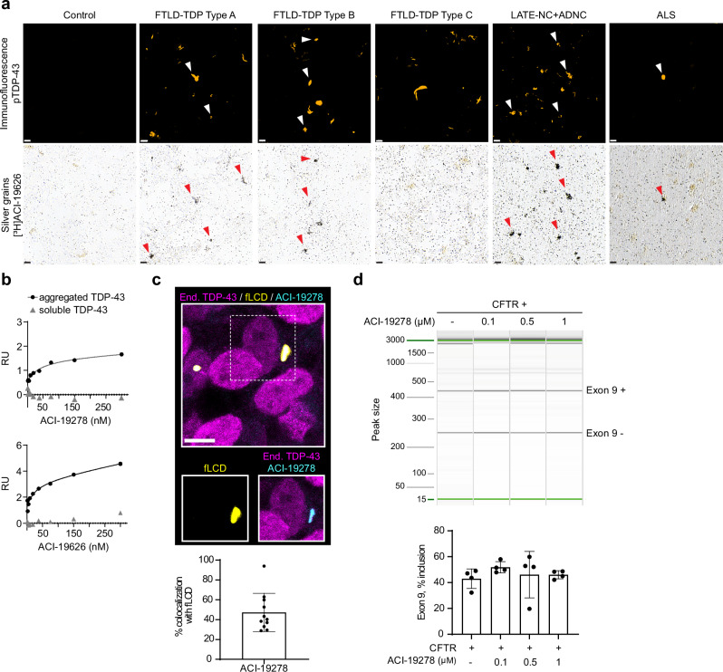
Aggregated TDP-43 is a hallmark of frontotemporal dementia (FTD), amyotrophic lateral sclerosis (ALS), and limbic-predominant age-related TDP-43 encephalopathy (LATE), and a common co-pathology in other neurodegenerative diseases. Currently, no specific biomarkers exist to assess TDP-43 pathology in vivo. We developed two small-molecule radiopharmaceuticals, [18F]ACI-19278 and [18F]ACI-19626, for visualizing TDP-43 inclusions by positron emission tomography (PET). Both ligands bind with high affinity to aggregated, but not soluble, TDP-43 in patient brain samples from diverse TDP-43 proteinopathies, including frontotemporal lobar degeneration with TDP-43 pathology (FTLD-TDP), ALS, and LATE, and in cell models. Both compounds display excellent selectivity for TDP-43 over Aβ, Tau, and α-synuclein aggregates. In non-human primates, [18F]ACI-19278 and [18F]ACI-19626 show a pharmacokinetic profile suitable for brain PET imaging (rapid brain uptake; fast and complete washout). ACI-19278 and ACI-19626 are promising first-in-class TDP-43 PET tracers with the potential to revolutionize the diagnosis and treatment of neurodegenerative proteinopathies, enabling a precision medicine approach.


Conflict of interest statement

Competing interests: EV is a consultant of AC Immune SA. EC, ND, DC, TM, JKB, MR, AMS, TJA, CD, MYR, HK, FCA, RLC, TA, AP, MKV and TS are or were employees of AC Immune SA at the time of their contribution to this manuscript. The remaining authors declare no competing interests.

Figures

Fig. 1
Fig. 1. Characterization of binding of [³H]ACI-19278 and [³H]ACI-19626 to brain samples from TDP-43 proteinopathies.
a Autoradiographic images of [3H]ACI-19278 and [3H]ACI-19626 binding in brain tissue sections from control (temporal cortex), FTLD-TDP (type A, frontal cortex; type B, temporal cortex), and LATE-NC + ADNC (hippocampus) cases. Total: total binding; Non-specific binding (NSB): residual binding in the presence of 2 μM unlabeled ACI-19278 or ACI-19626. Scale bar, 5 mm. Seven cases were tested for FTLD-TDP type A, five ([3H]ACI-19278) or seven ([3H]ACI-19626) cases for type B, and one case from LATE-NC + ADNC in independent experiments. b Specific binding (for ACI-19278: n = 9 control; n = 7 type A; n = 5 type B; n = 5 type C; n = 1 LATE-NC; n = 5 ALS. For ACI-19626: n = 7 control; n = 7 type A; n = 6 type B; n = 4 type C; n = 1 LATE-NC; n = 3 ALS). Data is shown as mean ± SD. One-way ANOVA with Dunnett’s multiple comparisons test; for ACI-19278 **p = 0.006 for control vs type A; for ACI-19626 *p = 0.01 for control vs type A, and **p = 0.009 for control vs type B. c Representative autoradiograms of [3H]ACI-19626 with higher magnification images of selected areas in adjacent sections following labeling with anti-phospho-TDP-43 antibody. Top row (images 1, 3, and 5), high-density TDP-43 pathology; bottom row (images 2, 4, 6), mid-low density TDP-43 pathology. Scale bar, 20 μm. d Saturation binding studies with [3H]ACI-19278 and [3H]ACI-19626 on human brain tissue sections from an FTLD-TDP type A donor. Data from one experiment. e Saturation binding studies with [3H]ACI-19278 and [3H]ACI-19626 in brain homogenates from control (n = 1), FTLD-TDP type A, type B, and type C cases. Data is shown as mean ± SD for [3H]ACI-19626 on type A (six independent experiments) and for both compounds on type C (three independent experiments). In other cases, representative data from two independent experiments is shown. f Kd values from saturation binding studies in e and Supplementary Fig. 1e. Mean ± SD is reported for FTLD-TDP type A and B across two cases. n.a., no fit or R2 < 0.85. Kd values in panels d and f were assessed by different techniques and on different tissue preparations. Source data are provided as a Source Data file.
Fig. 2
Fig. 2. Binding specificity of ACI-19278 and ACI-19626 to aggregated TDP-43.
a High-resolution autoradiography with [³H]ACI-19626 (20 nM) in tissue from FTLD-TDP type A, type B and type C, LATE-NC + ADNC, ALS cases and a healthy control. Immunofluorescence with phospho-TDP-43 pS409/410 (pTDP-43) antibody (top panels, white arrowheads). Accumulation of silver grains on pTDP-43 inclusions on the same section (bottom panels, red arrowheads), showing co-labeling of pTDP-43 aggregates. Scale bar, 10 μm. Representative images of two independent experiments and donors for each tissue type (except for LATE-ND + ADNC, 3 cases tested). b Surface plasmon resonance studies for the determination of binding affinity of ACI-19278 and ACI-19626 to aggregated and soluble TDP-43. Single-cycle kinetics analysis was performed on immobilized brain-derived TDP-43 aggregates from an FTLD-TDP type A donor and on full-length recombinant TDP-43. Response signals (RU) are plotted against ACI-19278 or ACI-19626 concentration and steady-state affinity fits to calculate Kd. Data from one out of four independent experiments are shown. c ACI-19278 efficiently binds to TDP-43 fibrils and not to nuclear physiological TDP-43. Top, representative immunofluorescence image showing SH-SY5Y cells treated with TDP-43 fibrils (fLCD, yellow) and endogenous TDP-43 (magenta) and with ACI-19278 (cyan). Scale bar, 5 μm. Bottom, quantification depicting the percentage of overlapping area of exogenous TDP-43 aggregates (fLCD) and ACI-19278. Data is shown as mean ± SD from two independent experiments (n = 11). d ACI-19278 does not interfere with the physiological function of TDP-43. RT-PCR amplification and capillary electrophoresis analysis of CFTR exon 9 inclusion in CTFR minigene transfected SH-SY5Y cells in the presence of ACI-19728 (0.1–1 µM). Results from four independent experiments) (bottom panel), data shown as mean ± SD. One-way ANOVA with Dunnett’s multiple comparisons test showed no statistically significant differences. Source data are provided as a Source Data file.
Fig. 3
Fig. 3. ACI-19278 and ACI-19626 selectivity for TDP-43 over common co-pathologies.
a Autoradiography in Alzheimer’s disease (AD) tissue containing amyloid and Tau aggregates with an Aβ reference compound ([³H]Aβ ref cmpd) and Tau reference ligand ([³H]PI-2620). NSB: non-specific binding. Immunolabeling of adjacent sections with phospho-TDP-43 (orange), phospho-Tau (green) or Aβ (red) antibody. Scale bar, 2 mm. Representative autoradiograms from one donor from 3 independent experiments. b High-resolution autoradiography in the entorhinal cortex sections of an AD patient. [3H]PI-2620 included for reference. Thioflavin S staining in an adjacent section (left, green). Scale bar, 50 µm. Representative data of two independent experiments. c High-resolution autoradiography in FTLD-Tau tissue sections. Immunolabeling with phospho-Tau (top panels, green). Scale bar, 50 µm. Representative data of two donors tested in one experiment. d High-resolution autoradiography in tissue sections from a LATE-NC + ADNC case. Immunolabeling of the same sections with phospho-TDP-43 (orange), phospho-Tau (green), or Aβ (red) antibodies. Autoradiography images from adjacent sections with [3H]PI-2620 and [3H]Aβ reference compounds are shown on the bottom. Scale bar, 20 µm. Representative data of two donors tested in one experiment. White arrowheads: pTDP-43 pathology; red arrowheads: compound binding. e Saturation binding studies with [³H]ACI-19278 and [³H]ACI-19626 in brain homogenates from: From left to right, AD brain tissue containing Aβ and Tau aggregates (AD, insoluble fraction), AD brain tissue enriched for insoluble Tau paired helical filaments (AD, Tau PHF), and Parkinson’s disease (PD) brain tissue enriched for α-synuclein aggregates (PD, α -syn enriched). Reference ligands specific for Aβ (Aβ ref cmpd), Tau (PI-2620, Tau ref cmpd), and α-synuclein (ACI-12589, α-syn ref cmpd), respectively, were assessed in each homogenate. Representative data of at least two independent experiments for [³H]ACI-19278 and [³H]ACI-19626, and mean ± SD of three independent experiments for the [³H]Aβ ref cmpd in AD brain homogenates. Data from one out of two donors tested in independent experiments are shown for PHF and PD brain homogenates. Dotted line shows the limit under which binding is considered not relevant. Source data are provided as a Source Data file.
Fig. 4
Fig. 4. Assessment of the pharmacokinetic profile of [18F]ACI-19278 and [18F]ACI-19626 as brain PET tracers in rhesus macaques.
The same rhesus macaque, receiving either [18F]ACI-19278 or [18F]ACI-19626 intravenously at different points in time, was imaged by PET. A magnetic resonance imaging (MRI) of the brain was acquired on a separate day for anatomical reference and image analysis. a Transverse, sagittal and coronal views for MRI and PET images are provided to illustrate the determination of standardized uptake values (SUV) averaged over 0–30 min (middle row) and 30–90 min post-injection (bottom row). b Time activity curves (TACs) generated from the data obtained by quantification of the SUV for either [18F]ACI-19278 or [18F]ACI-19626 from different brain regions. Data from one experiment. c Comparison of the TACs for [18F]ACI-19278 and [18F]ACI-19626 averaged across the whole brain and reported as a percentage of the highest uptake (SUVmax) for each molecule. Source data are provided as a Source Data file.

References

    1. Neumann, M. et al. Ubiquitinated TDP-43 in frontotemporal lobar degeneration and amyotrophic lateral sclerosis. Science314, 130–133 (2006). - PubMed
    1. Arai, T. et al. TDP-43 is a component of ubiquitin-positive tau-negative inclusions in frontotemporal lobar degeneration and amyotrophic lateral sclerosis. Biochem Biophys. Res. Commun.351, 602–611 (2006). - PubMed
    1. Nelson, P. T. et al. Limbic-predominant age-related TDP-43 encephalopathy (LATE): consensus working group report. Brain142, 1503–1527 (2019). - PMC - PubMed
    1. Uryu, K. et al. Concomitant TAR-DNA-binding protein 43 pathology is present in Alzheimer disease and corticobasal degeneration but not in other tauopathies. J. Neuropathol. Exp. Neurol.67, 555–564 (2008). - PMC - PubMed
    1. Arai, T. et al. Phosphorylated TDP-43 in Alzheimer’s disease and dementia with Lewy bodies. Acta Neuropathol.117, 125–136 (2009). - PubMed

MeSH terms

LinkOut - more resources