

Pars plana vitrectomy in progressive severe stellate non-hereditary idiopathic foveomacular retinoschisis (SNIFR): surgical outcomes and considerations for pathophysiology
- PMID: 41137096
- PMCID: PMC12553245
- DOI: 10.1186/s40942-025-00742-w
Pars plana vitrectomy in progressive severe stellate non-hereditary idiopathic foveomacular retinoschisis (SNIFR): surgical outcomes and considerations for pathophysiology
Abstract
Background: To report the clinical course and outcomes of a surgical approach for progressive severe stellate non-hereditary idiopathic foveomacular retinoschisis (SNIFR) using pars plana vitrectomy (PPV).
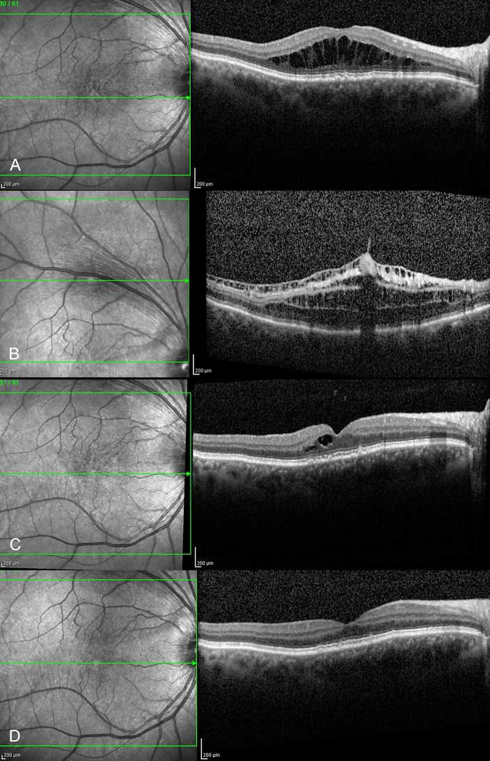
Methods: Multi-center, consecutive, interventional case series. Patients with a diagnosis of SNIFR presenting with progressive loss of vision between January 1, 2017 and January 1, 2023. Evaluation of ophthalmologic findings and multimodal ocular imaging at the time of diagnosis, surgical procedure, and of visual and anatomic outcomes postoperatively. The main outcome measures evaluated include best corrected visual acuity (BCVA), central macular thickness (CMT), and findings on optical coherence tomography (OCT).
Results: Seven patients diagnosed with SNIFR were included. Median age in years at the time of diagnosis was 64 (range, 46-77). Four patients were female, and three were male. Genetic testing for mutations in retinoschisin 1 (RS1) and for other inherited conditions associated with foveomacular retinoschisis was negative. All patients demonstrated progressive and severe retinoschisis, as well as worsening vision loss and metamorphopsia when managed conservatively. PPV was performed and revealed anomalously broad and dense adherence of the posterior hyaloid in all eyes. The internal limiting membrane (ILM) was peeled in all but one case. Median BCVA at baseline measured 20/50, and declined to 20/70 at the time of surgery. Median preoperative CMT measured 561 μm, with OCT demonstrating prominent retinoschisis of the outer plexiform and outer nuclear layers. All eyes demonstrated postoperative resolution of retinoschisis and subretinal fluid, with corresponding improvements in both BCVA and subjective central visual distortion up to six months after surgery. BCVA for the entire cohort improved to a median of 20/30, and with a corresponding decrease in CMT to a median of 240 μm.
Conclusion: PPV is an effective surgical intervention resulting in anatomic resolution of retinoschisis and improved functional vision in eyes with progressive and severe SNIFR.
Keywords: Foveomacular schisis; Hyaloid; Idiopathic; Internal limiting membrane; Macular thickness; Membrane peel; Pars plana vitrectomy; Retinoschisis.
© 2025. The Author(s).
Conflict of interest statement
Declarations. Ethics approval and consent to participate: Approved by the ethics committee/institutional review board at Oakland University William Beaumont School of Medicine, Rochester, Michigan. Consent for publication: Written informed consent for research and publication was obtained from all patients. Competing interests: The authors declare no competing interests.
Figures


References
LinkOut - more resources
Full Text Sources
Miscellaneous
