Case Reports
. 2025 Oct 16;21(1):224-228.
doi: 10.1016/j.radcr.2025.09.070. eCollection 2026 Jan.

Sickle cell disease presenting with intrahepatic extramedullary hematopoiesis: MRI characterization and diagnostic clues

Affiliations
Case Reports

Sickle cell disease presenting with intrahepatic extramedullary hematopoiesis: MRI characterization and diagnostic clues

Ayham Khan Ansari et al. Radiol Case Rep. .

Abstract

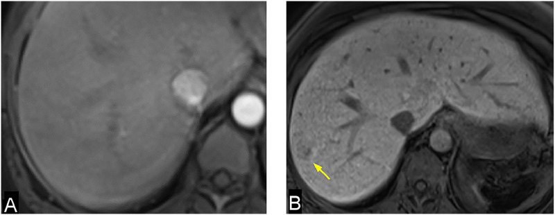
Extramedullary hematopoiesis (EMH) refers to the formation of hematopoietic tissue outside the bone marrow and usually occurs in response to chronic anemia or marrow dysfunction. While EMH is well-documented in conditions such as thalassemia and myeloproliferative disorders, intrahepatic extramedullary hematopoiesis is very rarely documented in sickle cell disease. We present the MRI imaging features of a rare case of intrahepatic extramedullary hematopoiesis in a patient with sickle cell disease. Given the potential for misdiagnosis as primary hepatic malignancy or as infectious lesions, this case report underscores the importance of recognizing the imaging features of intrahepatic extramedullary hematopoiesis in the context of sickle cell disease and chronic transfusion-related iron overload.

Keywords: Extramedullary hematopoiesis; Intrahepatic lesions; MRI; Sickle cell disease.


Figures

Fig 1
Fig. 1
Axial T2 WI (A) shows diffuse low signal intensity of the liver parenchyma indicative of iron deposition. A mildly hyperintense lesion is seen in segment VIII (yellow arrow). Also note the small sized spleen with diffuse low signal intensity due to iron deposition and calcifications. Axial T2 FS images (B & C) show other smaller hyperintense lesions in the right lobe at segments VII, V and VI (yellow arrows).
Fig 2
Fig. 2
Axial DWI (A) and ADC map (B) show T2 shine through of the lesion without diffusion restriction appearing as hyperintense signal in DWI and ADC map (yellow arrows). All other lesions showed similar signal intensity (not shown).
Fig 3
Fig. 3
Axial T1 FS arterial phase (A) shows isointense signal of the lesion to the liver parenchyma without appreciable enhancement. Axial delayed 20 minutes T1 FS Dixon hepatobiliary phase shows hypointensity of the lesion relative to the surrounding liver parenchyma. Also note diffuse reticular pattern of the liver parenchyma in the delayed hepatobiliary phase, reflecting chronic parenchymal disease. All other lesions showed similar signal intensity (not shown).
Fig 4
Fig. 4
Percutaneous ultrasound guided biopsy, needle tip is seen within a peripheral hypoechoic lesion at segment VIII.
Fig 5
Fig. 5
The histologic sections of the liver cores biopsy. (A&B) Hematoxylin and eosin (H&E) staining show benign liver parenchyma with mildly dilated sinusoids, occasional mature megakaryocytes and erythroid precursors. Immunohistochemical stains for CD61 (C) highlighting megakaryocytes and Glycophorin C (D) in erythroid elements. Original magnification x100 (A), x400 (B), x200 (C&D).

References

    1. Cenariu D, Iluta S, Zimta A-A, Petrushev B, Qian L, Dirzu N, et al. Extramedullary hematopoiesis of the liver and spleen. J Clin Med. 2021;10(24):5831. doi: 10.3390/jcm10245831. - DOI - PMC - PubMed
    1. Ajayi F., Nali M.S., Ali R., Patel A., Shaaban H. Extra-medullary hematopoiesis in sickle cell disease presenting as a right adrenal mass. Cureus. 2022;14(1) doi: 10.7759/cureus.21334. - DOI - PMC - PubMed
    1. Jelali MA, Luciani A, Kobeiter H, Zafrani S, Anglade MC, Zegai B, et al. MRI features of intrahepatic extramedullary haematopoiesis in sickle cell anaemia. Cancer Imaging. 2006;6(1):182–185. doi: 10.1102/1470-7330.2006.0030. - DOI - PMC - PubMed
    1. La Fianza A., van der Byl G., Maccabelli G., Torretta L., Calliada F. CT and MR findings in extramedullary haematopoiesis with biliary system encasement: a case report. J Radiol Case Rep. 2010;4(11):1–8. doi: 10.3941/jrcr.v4i11.462. - DOI - PMC - PubMed
    1. Wong Y, Chen F, Tai KS, Yip LK, Tsang KW, Chan FL, et al. Imaging features of focal intrahepatic extramedullary haematopoiesis. Br J Radiol. 1999;72(861):906–910. doi: 10.1259/bjr.72.861.10645201. - DOI - PubMed

Publication types

LinkOut - more resources