. 2025 Oct 27;30(1):123.
doi: 10.1186/s11658-025-00800-z.

KIAA1429-mediated M6A methylation inhibits osteoclast differentiation via stabilizing Lrp4 mRNA and protects against osteoporosis

Affiliations

KIAA1429-mediated M6A methylation inhibits osteoclast differentiation via stabilizing Lrp4 mRNA and protects against osteoporosis

Jincheng Liu et al. Cell Mol Biol Lett. .

Abstract

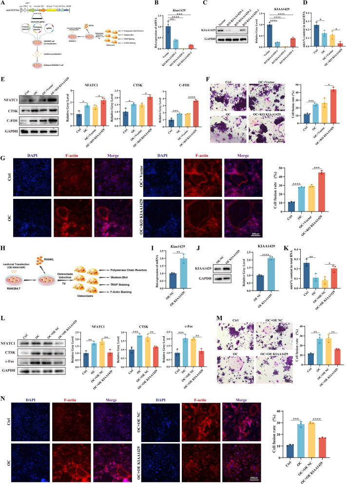
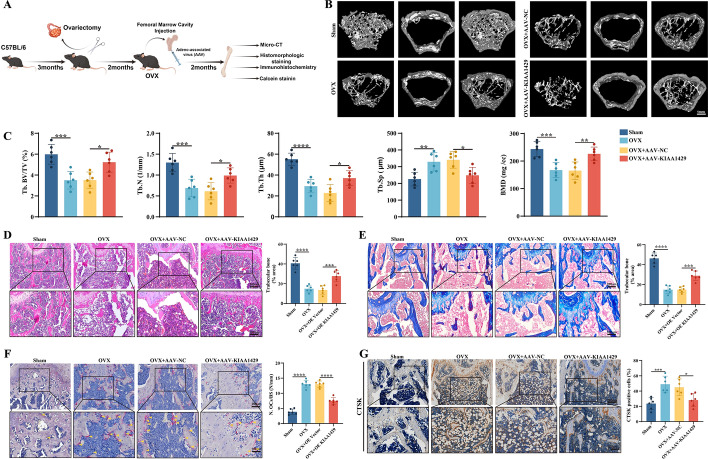
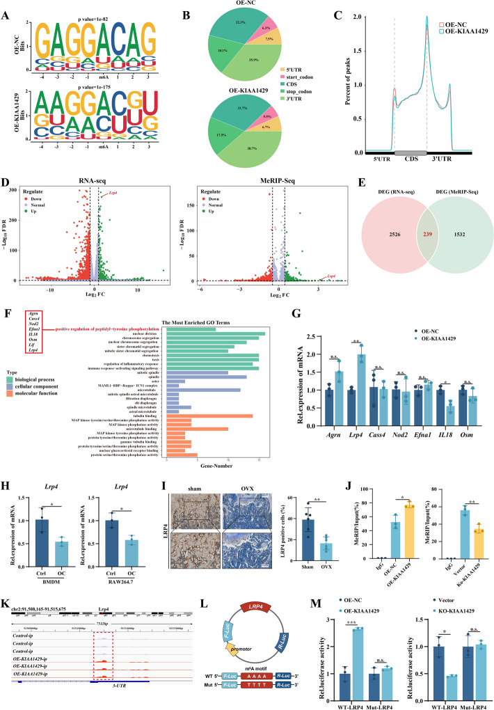
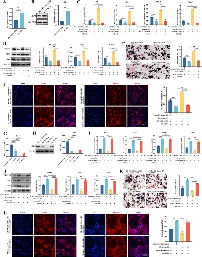
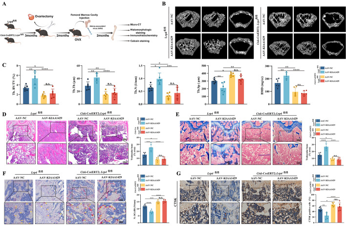
N6-methyladenosine (m6A) is a novel epigenetic modification that has been reported to be involved in the progression of osteoporosis (OP), providing new insights into the pathogenesis of OP. The methyltransferases KIAA1429 [also known as virus-like m6A methyltransferase-associated protein (VIRMA)] participates in various essential biological processes by regulating target gene expression levels. However, the function of KIAA1429-mediated m6A modification in OP progression remains unclear. This study aimed to investigate the biological roles and potential underlying mechanisms of KIAA1429 in OP and osteoclast differentiation. scRNA-seq combined with bulk RNA-seq screening for the differential gene KIAA1429. Analysis of clinical data confirmed KIAA1429 expression and its clinical significance in OP. KIAA1429 inhibited osteoclast differentiation in vitro and reduced bone resorption in ovariectomized (OVX) mice. Mechanistically, LRP4 was identified as a downstream target of KIAA1429. KIAA1429 mediated the m6A modification of Lrp4 mRNA, and then YT521-B homology-domain-containing protein 1 (YTHDC1) increased Lrp4 stability and expression. In addition, LRP4 enhancement recruited TNFAIP3, which inactivated NF-κB signaling. This novel mechanism of NF-κB signaling pathway inhibition by enhanced KIAA1429/YTHDC1-coupled Lrp4 transcription during osteoclast differentiation highlights the potential of KIAA1429 as a novel predictive biomarker and therapeutic target for OP progression.

Keywords: N 6-methyladenosine; KIAA1429; LRP4; Osteoclast; Osteoporosis.


Conflict of interest statement

Declarations. Ethics approval and consent to participate: Human subjects research was approved by the Medical Ethics Committee of Qilu Hospital (Qingdao) of Shandong University (approval no. KYLL-2024036, date: 28 October 2024) and performed in accordance with the principles of the Declaration of Helsinki. Informed consent was obtained from all subjects involved in the study. All animal studies were approved by the Animal Care and Ethics Committee of Shandong University (approval no. 24015, date: 24 April 2024). The Animal Care and Ethics Committee of Shandong University adheres to the principles and guidelines set forth by the International Council for Laboratory Animal Science (ICLAS), ensuring that our animal research meets international ethical standards. Consent for publication: Not applicable. Competing Interests: The authors declare no competing interests.

Figures

Fig. 1
Fig. 1
KIAA1429 and m6A modification were decreased in osteoporotic bone tissues and were closely associated with osteoclast differentiation. A Heatmap of m6A regulator expression in the peripheral blood mononuclear cell (PBMC) and osteoclast (OC) from the GEO database (GSE225974), with high and low expression levels shown in red and blue, respectively. B Schematic workflow of the scRNA-seq. C A UMAP analysis results displaying 18 cell clusters. D Annotation of 18 cell clusters to eight cell types. E Volcano plots of different cellular subset in sham and OVX mice in scRNA-seq. F UMAP expression distribution of specific marker genes for osteoclasts and Kiaa1429. G Violin plot demonstrating the difference in Kiaa1429 expression in osteoclasts between sham and OVX mice. H Representative images of tartrate-resistant acid phosphatase (TRAP) staining of RAW264.7 cells on days 0, 3, and 5 during the osteoclast differentiation. I mRNA levels of osteoclast-related genes (Ctsk, c-Fos, and Mmp-9) in RAW264.7 on days 0, 3, and 5 during the osteoclast differentiation were assayed by qRT-PCR analysis. J mRNA level of Kiaa1429 in RAW264.7 on days 0, 3, and 5 during the osteoclast differentiation was assayed by qRT-PCR analysis. K The m6A content from RAW264.7 was measured by colorimetric quantification assay on days 0, 3, and 5 during the osteoclast differentiation. L Representative images of TRAP staining in primary bone marrow-derived macrophages (BMDM) on days 0, 3, and 5 during the osteoclast differentiation. M mRNA levels of osteoclast-related genes in BMDM on days 0, 3, and 5 during the osteoclast differentiation were assayed by qRT-PCR analysis. N mRNA level of Kiaa1429 in BMDM on days 0, 3, and 5 during the osteoclast differentiation was assayed by qRT-PCR analysis. O The m6A content from BMDM was measured by colorimetric quantification assay on days 0, 3, and 5 during the osteoclast differentiation. P mRNA level of Kiaa1429 in bone tissues of sham and OVX mice was assayed by qRT-PCR analysis. Q The m6A content in bone tissues of sham and OVX mice was measured by colorimetric quantification assay. R Representative images of immunohistochemistry staining for KIAA1429 in sham and OVX mice. S The serum concentration of CTX-1 in sham and OVX mice. T Co-expression analysis of Kiaa1429 mRNA expression with CTX-1 in sham and OVX mice. U mRNA level of KIAA1429 in bone tissues of postmenopausal patients with osteoporosis and patients without osteoporosis was assayed by qRT-PCR analysis. V The m6A content in bone tissues of patients with osteoporosis and patients without osteoporosis was measured by colorimetric quantification assay. W The serum concentration of CTX-1 in postmenopausal patients with osteoporosis and patients without osteoporosis. X Co-expression analysis of the KIAA1429 mRNA with CTX-1 in postmenopausal patients with osteoporosis and patients without osteoporosis. Y Co-expression analysis of the KIAA1429 mRNA with L1–L4 BMD T-scores or hip BMD T-scores in postmenopausal patients with osteoporosis and patients without osteoporosis. The values are mean ± SD of at least three independent experiments; n.s. not significant; n.s.p > 0.05, *p < 0.05, **p < 0.01, ***p < 0.001, ****p < 0.0001
Fig. 2
Fig. 2
KIAA1429 knockout promoted osteoclast differentiation and KIAA1429 overexpression inhibited osteoclast differentiation in vitro. A Workflow of generating Kiaa1429 knockout RAW264.7 cells using CRISPR/Cas9 technology and subsequent osteoclast differentiation related-tests. B, C KIAA1429 expression in vector and Kiaa1429 knockout RAW264.7 cells, measured by qRT-PCR and western blotting. D The m6A content was measured in Kiaa1429 knockout cells. E Immunoblotting of osteoclast-related proteins (NFATC1, CTSK, and C-FOS) in control and Kiaa1429 knockout cells with or without RANKL treatment. F, G Representative images of TRAP staining and F-actin staining in Kiaa1429 knockout cells. H Workflow of osteoclast differentiation related-tests in control and Kiaa1429-overexpressed cell. I, J KIAA1429 expression in Kiaa1429-overexpressed RAW264.7 cells, measured by qRT-PCR and western blotting. K The m6A content in Kiaa1429-overexpressed cells was measured by colorimetric quantification assay. L Immunoblotting of osteoclast-related proteins (NFATC1, CTSK, and C-FOS) in control and Kiaa1429-overexpressed cells with or without RANKL treatment. (M, N) Representative images of TRAP staining and F-actin staining in Kiaa1429-overexpressed cells. The values are mean ± SD of at least three independent experiments; n.s.p > 0.05, *p < 0.05, **p < 0.01, ***p < 0.001, ****p < 0.0001
Fig. 3
Fig. 3
Kiaa1429 overexpression inhibited bone resorption in vivo. A Flowchart of the AAV injection experiment into the bone marrow cavity. B Representative micro-CT images of trabecular bone from the femoral metaphysis. C Micro-CT measurements of Tb. BV/TV; Tb. Th; Tb. No.; Tb. Sp; and BMD in the femurs after treatment with overexpressed negative control (AAV-NC) and overexpressed Kiaa1429 adeno-associated virus (AAV-KIAA1429). D Representative images of H&E staining in AAV-NC and AAV-KIAA1429 groups. E Representative images of Masson staining in AAV-NC and AAV-KIAA1429 groups. F Representative images of TRAP staining in AAV-NC and AAV-KIAA1429 groups. G Representative images of immunohistochemistry staining for CTSK in AAV-NC and AAV-KIAA1429 groups. The values are mean ± SD of at least three independent experiments; n.s.p > 0.05, *p < 0.05, **p < 0.01, ***p < 0.001, ****p < 0.0001
Fig. 4
Fig. 4
LRP4 was identified as a downstream target of KIAA1429-mediated m6A modification in osteoclast differentiation. A HOMER with MeRIP-seq identified the top consensus motif of m6A peaks in RAW264.7 with or without Kiaa1429 overexpression. B, C The percentage of total m6A peak distribution in the specified regions in RAW264.7 cells with OE NC and KIAA1429 overexpression. D Volcano plot of RNA-seq and MeRIP-seq in OE NC and Kiaa1429-overexpressed cells. E Venn diagram exhibiting overlap among differentially expressed genes (DEGs) on the basis of MeRIP-seq and RNA-seq. F Gene ontology (GO) enrichment analysis revealed high enrichment of the positive regulation of peptidyl-tyrosine phosphorylation pathway on the basis of RNA-seq and MeRIP-seq. G mRNA expression of DEGs upon Kiaa1429 overexpression, measured by qRT-PCR. H mRNA level of Lrp4 in RAW264.7 and BMDM during osteoclast differentiation, measured by qRT-PCR. I Representative images of immunohistochemistry staining for LRP4 in sham and OVX mice and quantitative analysis. J MeRIP-qPCR demonstrated the direct binding between m6A and Lrp4 mRNA. K Visualization of m6A peak in Lrp4 transcripts based on MeRIP-seq. L Construction of a luciferase reporter containing wild-type (WT) Lrp4 3′-UTR and mutation (Mut) Lrp4 3′-UTR at the m6A consensus sequence. M Luciferase activity in Kiaa1429 knockout or Kiaa1429-overexpressed RAW264.7 cells. The values are mean ± SD of at least three independent experiments; n.s.p > 0.05, *p < 0.05, **p < 0.01, ***p < 0.001, ****p < 0.0001
Fig. 5
Fig. 5
LRP4 overexpression rescued the osteoclast differentiation induced by Kiaa1429 knockout, and LRP4 knockout reversed the osteoclast differentiation induced by Kiaa1429 overexpression. A, B LRP4 expression in Lrp4-overexpressed RAW264.7 cells, assayed by qRT-PCR and western blotting. C mRNA expressions of Ctsk, C-fos, Nfatc1, and Mmp9 in Kiaa1429 knockout cells with or without Lrp4 overexpression after RANKL treatment. D Immunoblotting of osteoclast-related proteins (NFATC1, CTSK, and C-FOS) in Kiaa1429 knockout cells with or without Lrp4 overexpression after RANKL treatment. The vector group was used as the control group for KO-KIAA1429, and NC group was used as the control group for OE-LRP4. (E, F) TRAP staining and F-actin in Kiaa1429 knockout cells with or without Lrp4 overexpression after RANKL treatment. G, H qRT-PCR and western blotting confirmation of Lrp4 knockout in RAW264.7 cells. I, J The effect of Lrp4 knockout on osteoclast differentiation was measured by qRT-PCR and western blotting after Kiaa1429 overexpression. K, L TRAP staining and F-actin in Lrp4 knockout cells with or without Kiaa1429 overexpression after RANKL treatment. The values are mean ± SD of at least three independent experiments; n.s.p > 0.05, *p < 0.05, **p < 0.01, ***p < 0.001, ****p < 0.0001
Fig. 6
Fig. 6
Kiaa1429 overexpression could not rescue bone loss by LRP4 deficiency in vivo. A Flowchart of the AAV injection experiment into the bone marrow cavity in Ctsk-CreERT2; Lrp4fl/fl OVX mice and Lrp4fl/fl OVX mice. B Representative micro-CT images of trabecular bone from the femoral metaphysis in Ctsk-CreERT2; Lrp4fl/fl OVX mice and those of other groups. C Micro-CT measurements of Tb. BV/TV; Tb. Th; Tb. No.; Tb. Sp; and BMD in the femurs of Ctsk-CreERT2; Lrp4fl/fl OVX mice after treatment with AAV-NC or AAV-KIAA1429. D Representative images of H&E staining and quantitative analysis in Ctsk-CreERT2; Lrp4fl/fl OVX mice and Lrp4fl/fl OVX mice after treatment with AAV-NC or AAV-KIAA1429. E Representative images of Masson staining and quantitative analysis in Ctsk-CreERT2; Lrp4fl/fl OVX mice and Lrp4fl/fl OVX mice after treatment with AAV-NC or AAV-KIAA1429. F Representative images of TRAP staining and quantitative analysis in Ctsk-CreERT2; Lrp4fl/fl OVX mice and Lrp4fl/fl OVX mice after treatment with AAV-NC or AAV-KIAA1429. G Representative images of immunohistochemistry staining for CTSK and quantitative analysis in Ctsk-CreERT2; Lrp4fl/fl OVX mice, and Lrp4fl/fl OVX mice after treatment with AAV-NC or AAV-KIAA1429. The values are mean ± SD of at least three independent experiments; n.s.p > 0.05, *p < 0.05, **p < 0.01, ***p < 0.001, ****p < 0.0001
Fig. 7
Fig. 7
KIAA1429 improved Lrp4 mRNA stability through a YTHDC1-mediated methylation recognition. A Heatmap of m6A readers expression in RNA-seq, with high and low expression levels shown in red and blue, respectively. B qRT-PCR was used to determine the alterations in mRNA expressions of m6A readers upon Kiaa1429 overexpression. C Validation of direct binding between YTHDC1 and Lrp4 mRNA was performed by RIP-qPCR. D, E qRT-PCR and western blotting confirmation of Ythdc1 knockdown RAW264.7 cells. (F, G, H) LRP4 expression was measured by qRT-PCR and western blotting in Kiaa1429 overexpression cells after treatment with si-YTHDC1. (I) mRNA expressions of Ctsk, C-fos, Nfatc1 and Mmp9 in Kiaa1429 overexpression cells after treatment with si-YTHDC J, K Representative images of TRAP and F-actin staining and quantitative analysis in Kiaa1429 overexpression cells after treatment with si-YTHDC1. L Flow chart of the increased stability of Lrp4 mRNA in a YTHDC1-dependent manner. The values are mean ± SD of at least three independent experiments; n.s.p > 0.05, *p < 0.05, **p < 0.01, ***p < 0.001, ****p < 0.0001
Fig. 8
Fig. 8
KIAA1429-mediated LRP4-regulated TNFAIP3 in osteoclasts. A Kyoto Encyclopedia of Genes and Genome (KEGG) enrichment analysis of signaling pathway based on RNA-seq and MeRIP-seq. B qRT-PCR was used to determine the alterations in mRNA expression of DEGs upon Kiaa1429 overexpression. C Western blotting was performed for TNFAIP3 in Kiaa1429-overexpressed RAW264.7 cells. D, E qRT-PCR and western blotting were performed for TNFAIP3 in Lrp4-overexpressed cells. F Representative images of immunohistochemistry staining for TNFAIP3 and quantitative analysis in Ctsk-CreERT2; Lrp4fl/fl and Lrp4fl/fl OVX mice. G, H TNFAIP3 was measured by qRT-PCR and western blotting in RAW264.7 and BMDM during osteoclast differentiation. I, J The effect of Lrp4 knockout on TNFAIP3 expression was measured by qRT-PCR and western blotting after Kiaa1429 overexpression. K, L, M The effect of Lrp4 overexpression on TNFAIP3 expression was measured by qRT-PCR and western blotting after Kiaa1429 knockout. N Co-immunoprecipitation of TNFAIP3 with endogenous LRP4 in RAW264.7 and BMDM. O Co-localization of LRP4 and TNFAIP3 after treatment with or without RANKL in RAW264.7 cells was exhibited in confocal immunofluorescent images. The values are mean ± SD of at least three independent experiments; n.s.p > 0.05, *p < 0.05, **p < 0.01, ***p < 0.001, ****p < 0.0001
Fig. 9
Fig. 9
KIAA1429/LRP4-mediated TNFAIP3 inhibited osteoclast differentiation by activating the NF-κB pathway. A, B TNFAIP3 expression was measured by qRT-PCR and western blotting in Tnfaip3 knockout RAW264.7 cells. C mRNA expressions of Ctsk, C-fos, Nfatc1, and Mmp9 in Tnfaip3 knockout cells with or without Lrp4 overexpression after RANKL treatment. D Immunoblotting of osteoclast-related proteins (NFATC1, CTSK, and C-FOS) in Tnfaip3 knockout cells with or without Lrp4 overexpression after RANKL treatment. E, F TRAP staining and fibrous actin (F-actin) staining in Tnfaip3 knockout cells with or without Lrp4 overexpression after RANKL treatment. G, H The activation of the NF-κB pathway (Phospho-p65, p65, Phospho-lkBα, lkBa, Phospho-IKKα/β, and IKKα) was measured by western blotting in Tnfaip3 knockout RAW264.7 cells with or without Lrp4 overexpression. I Expression of p65 in cytoplasmic or nuclear extracts was detected using western blotting in Tnfaip3 knockout RAW264.7 cells with or without Lrp4 overexpression. GAPDH and Lamin B were shown as cytoplasmic and nuclear internal controls, respectively. The values are mean ± SD of at least three independent experiments; n.s.p > 0.05, *p < 0.05, **p < 0.01, ***p < 0.001, ****p < 0.0001
Fig. 10
Fig. 10
KIAA1429-mediated m6A methylation inhibits osteoclast differentiation and protects against osteoporosis. KIAA1429 mediates m6A modification of Lrp4 mRNA, and then recruits YTHDC1 to increase Lrp4 stability and expression. Enhancement of LRP4 recruits TNFAIP3, leading to inactivation of NF-κB signaling, which in turn inhibits osteoclast differentiation

References

    1. Ensrud KE, Crandall CJ. Osteoporosis. Ann Intern Med. 2017;167(3):Itc17-itc32. - PubMed
    1. Li J, Chen X, Lu L, Yu X. The relationship between bone marrow adipose tissue and bone metabolism in postmenopausal osteoporosis. Cytokine Growth Factor Rev. 2020;52:88–98. - PubMed
    1. Raisz LG. Pathogenesis of osteoporosis: concepts, conflicts, and prospects. J Clin Invest. 2005;115(12):3318–25. - PMC - PubMed
    1. Subarajan P, Arceo-Mendoza RM, Camacho PM. Postmenopausal osteoporosis: a review of latest guidelines. Endocrinol Metab Clin North Am. 2024;53(4):497–512. - PubMed
    1. Compston JE, McClung MR, Leslie WD. Osteoporosis. Lancet. 2019;393(10169):364–76. - PubMed

MeSH terms