. 2025 Oct 13:36:100851.
doi: 10.1016/j.phro.2025.100851. eCollection 2025 Oct.

Apparent diffusion coefficient as a quantitative biomarker for prostate cancer treatment response on a 1.5 Tesla magnetic resonance-linear accelerator: Impact of image registration and acquisition type

Affiliations

Apparent diffusion coefficient as a quantitative biomarker for prostate cancer treatment response on a 1.5 Tesla magnetic resonance-linear accelerator: Impact of image registration and acquisition type

Prashant P Nair et al. Phys Imaging Radiat Oncol. .

Abstract

Background and purpose: Diffusion-weighted magnetic resonance imaging (DW-MRI) is a quantitative biomarker for cancer detection and treatment monitoring. On magnetic resonance-linear accelerator (MR-Linac) systems, diffusion-weighted echo planar imaging (DW-EPI) suffers from geometric distortion, reducing the repeatability of apparent diffusion coefficient (ADC) measurements. This study evaluated the effect of low-distortion split acquisition of fast spin-echo signal (SPLICE) sequences, and of image registration on the repeatability coefficient (RC) of ADC.

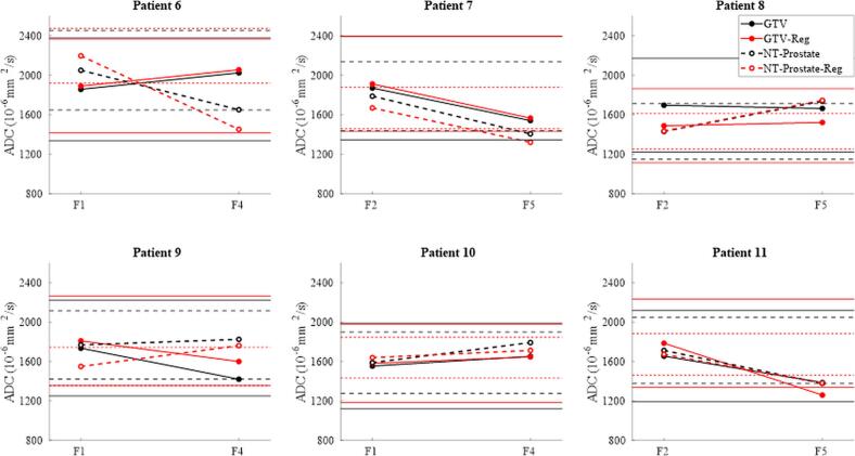
Materials and methods: ADC bias, repeatability, signal-to-noise ratio (SNR) and geometric fidelity were measured in a diffusion phantom using three DW-EPI and two DW-SPLICE protocols. ADC short-term and long-term RCs were measured in healthy volunteers. In patients, the registration of DW-EPI to unweighted images (b0) was tested for its effect on RC in gross tumour volume (GTV) and non-tumour prostate (NT-P), and for its ability to detect significant ADC changes.

Results: Phantom experiments showed strong linear correlation with ground-truth ADC (R2 > 0.99). Among EPI protocols, DW-EPI-AP offered the best balance of high SNR and low RC, while Z-direction encoded DW-EPI was the most variable. Both DW-SPLICE variants exhibited reduced distortion compared with EPI but poorer repeatability. In volunteers, long-term RCs (8.0-33.7 %) varied more than short-term RCs (8.9-15.4 %). In patients, registration improved RCs (GTV: 28.0 → 25.1 %; NT-P: 19.6 → 12.6 %) and improved detection of significant ADC change in patients (GTV: 0/6 → 1/6; NT-P: 2/6 → 5/6).

Conclusion: RC and accuracy of DW-EPI agrees with published literature and improves after registration. DW-SPLICE shows lower geometric distortion but would require further optimization and validation to improve repeatability.

Keywords: Apparent diffusion coefficient; Diffusion weighted echo planar imaging; Geometric distortion correction; MR-Linac; Repeatability coefficient.


Conflict of interest statement

The authors declare the following financial interests/personal relationships which may be considered as potential competing interests: PPN, JC, MN, BL, SC, ACT, PJvH, UO and AW declare the following conflict: The Institute of Cancer Research (ICR), the Royal Marsden Hospital (RMH) and the Netherlands Cancer Institute (NKI) are members of the MR-Linac Consortium with industrial partners Elekta and Philips. ICR and RMH receive research support from Elekta and Philips. ACT receives research funding from Elekta, Varian and Accuray, honoraria/travel assistance from Elekta, Accuray, Bayer and Janssen. ACT is chair of the MR linac consortium steering committee.

Figures

None
Graphical abstract
Fig. 1
Fig. 1
Linear fits of measured versus reference ADC values for each sequence are shown in 10−3 mm2/s, with R2, slope (proportional bias; ideal = 1), and intercept (additive bias; ideal = 0) reported alongside ±95 % CIs. All sequences exhibit strong linearity (R2). DW-EPI and DW-EPI-AP show no detectable bias. DW-EPI-Z demonstrates proportional but not additive bias. DW-SPLICE performs the worst, exhibiting both additive and proportional biases. EPI-b500 refers to ADC calculated the b-values of 0 and 500 s/mm2, unlike the others where ADC was derived using all the b-values.
Fig. 2
Fig. 2
Distances between markers in the phantom measured on various DW images and 3D T2W images, which served as distortion-free reference. All DW-EPI sequences caused significant distortion compared to the reference for all the marker distances, DW-SPLICE-LH showed reduced distortion, while DW-SPLICE showed no significant distortion compared to the T2. Values are represented as mean ± SD; * indicates p < 0.05; ** indicates p < 0.01.
Fig. 3
Fig. 3
Delta ADC measured in the prostate of three healthy volunteers for different DW sequences (colour-coded). Hollow markers denote short-term repeatability (within the same session). Filled markers denote long-term repeatability between sessions A and B (one week apart) across DW sequences for the three volunteers. Average delta ADC and ±1 SD are shown. The bar graphs show ADC values across sequences. The EPI-Z sequence exhibits significantly higher ADCs compared to all other sequences. DW-SPLICE-LH shows significantly higher ADCs than DW-EPI and DW-SPLICE, between which no significant difference is observed.
Fig. 4
Fig. 4
Registration results for a slice containing the GTV anatomy from patient P11. The non-deformed b0 image (fixed) is shown along with deformation fields in the left–right (LR) and anterior–posterior (AP) directions for each b-value and diffusion encoding direction, overlaid on the corresponding diffusion image (moving images). Numbers indicate the average deformation (mm) within the prostate contour. For each diffusion-weighting direction, the scanner’s three physical axes (X, Y, Z) contribute: Dir 1 = [−0.67, 0.33, −0.67], Dir 2 = [0.33, −0.67, −0.67] and Dir 3 = [−0.67, −0.67, 0.33]. Globally, deformation is higher for higher b-values. Deformations for diffusion direction 2 follows an opposite pattern to that of the deformations in diffusion encoding 1 and 3.
Fig. 5
Fig. 5
Mean ADC for the GTV, non-tumour prostate (NT-P) and whole prostate (WP) in the 14 scans from the 7 fractions used to calculate the RC. Registration reduced the ADC in the whole prostate. The ADC in the GTV is significantly lower than for NT-P for both the registered (Reg) and the non-registered (non-Reg) workflows.
Fig. 6
Fig. 6
ADC measurements for six patients in the GTV and non-tumour prostate (NT-P) from the five-fraction cohort, comparing non-registered (black) and registered (red) data. Measurements were taken at two different fractions (F), roughly one week apart. Dashed lines and hollow circles represent NT-P while bold lines and filled circles represent GTV. Horizontal lines represent 95% confidence limits for detecting a true change. (For interpretation of the references to colour in this figure legend, the reader is referred to the web version of this article.)

References

    1. Kim C.K., Park B.K., Kim B. Diffusion-weighted MRI at 3 T for the evaluation of prostate cancer. AJR Am J Roentgenol. 2010;194(6):1461–1469. doi: 10.2214/AJR.09.3654. - DOI - PubMed
    1. Koh D.M., Collins D.J. Diffusion-weighted MRI in the body: applications and challenges in oncology. AJR Am J Roentgenol. 2007;188(6):1622–1635. doi: 10.2214/AJR.06.1403. - DOI - PubMed
    1. Messina C., Bignone R., Bruno A., Bruno A., Bruno F., Calandri M., et al. Diffusion-weighted imaging in oncology: an update. Cancers (Basel) 2020;12(6) doi: 10.3390/cancers12061493. - DOI - PMC - PubMed
    1. Verma S., Rajesh A., Morales H., Lemen L., Bills G., Delworth M., et al. Assessment of aggressiveness of prostate cancer: correlation of apparent diffusion coefficient with histologic grade after radical prostatectomy. AJR Am J Roentgenol. 2011;196(2):374–381. doi: 10.2214/AJR.10.4441. - DOI - PubMed
    1. Zelhof B., Pickles M., Liney G., Gibbs P., Rodrigues G., Kraus S., et al. Correlation of diffusion-weighted magnetic resonance data with cellularity in prostate cancer. BJU Int. 2009;103(7):883–888. doi: 10.1111/j.1464-410X.2008.08130.x. - DOI - PubMed

LinkOut - more resources