. 2025 Oct 10;16(10):379.
doi: 10.3390/jfb16100379.

Short Expandable-Wing Suture Anchor for Osteoporotic and Small Bone Fixation: Validation in a 3D-Printed Coracoclavicular Reconstruction Model

Affiliations

Short Expandable-Wing Suture Anchor for Osteoporotic and Small Bone Fixation: Validation in a 3D-Printed Coracoclavicular Reconstruction Model

Chia-Hung Tsai et al. J Funct Biomater. .

Abstract

Suture anchors are widely used for tendon and ligament repair, but their fixation strength is compromised in osteoporotic bone and limited bone volume such as the coracoid process. Existing designs are prone to penetration and insufficient cortical engagement under such conditions. In this study, we developed a novel short expandable-wing (SEW) suture anchor (Ti6Al4V) designed to enhance pull-out resistance through a deployable wing mechanism that locks directly against the cortical bone. Finite element analysis based on CT-derived bone material properties demonstrated reduced intra-bone displacement and improved load transfer with the SEW compared to conventional anchors. Mechanical testing using matched artificial bone surrogates (N = 3 per group) demonstrated significantly higher static pull-out strength in both normal (581 N) and osteoporotic bone (377 N) relative to controls (p < 0.05). Although the sample size was limited, results were consistent and statistically significant. After cyclic loading, SEW anchor fixation strength increased by 25-56%. In a 3D-printed anatomical coracoclavicular ligament reconstruction model, the SEW anchor provided nearly double the fixation strength of the hook plate, underscoring its superior stability under high-demand clinical conditions. This straightforward implantation protocol-requiring only a 5 mm drill hole without tapping, followed by direct insertion and knob-driven wing deployment-facilitates seamless integration into existing surgical workflows. Overall, the SEW anchor addresses key limitations of existing anchor designs in small bone volume and osteoporotic environments, demonstrating strong potential for clinical translation.

Keywords: 3D-printing; coracoclavicular; osteoporotic; pull-out; suture anchor.


Conflict of interest statement

The authors declare no conflict of interest in this research.

Figures

Figure 1
Figure 1
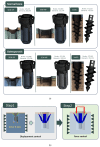
Design of the SEW anchor and instrument. Top left: dimensional drawing of the SEW anchor; bottom left: components including compressive head, four-blade body, and interlocking internal screw; center: instrument schematic; top right: knob and sleeve head grooves; middle right: square head of the sleeve with operation schematic; bottom right: triangular head of the internal screw with operation schematic.
Figure 2
Figure 2
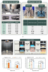
Compression testing of artificial bone blocks with different densities. Representative images of 20 PCF solid, 15 PCF cellular, 7.5 PCF cellular, and 7.5 PCF open foam blocks (top). Schematic of compression setup with a load cell at 5 mm/min (bottom left). Average elastic modulus (E), standard deviation (SD), and sample number (N = 3) for each group are summarized in the table (bottom right).
Figure 3
Figure 3
Finite element analysis (FEA) setup for SEW and control anchors. (a) Mesh models of SEW-OP, SEW-NP, and Smith anchors in normal and osteoporotic bone conditions, showing corresponding node and element counts. (b) Two-stage simulation process: Step 1—displacement control for wing expansion (1.9 mm in normal bone or 3.0 mm in osteoporotic bone); Step 2—force control with an upward pull-out load of 400 N applied to the anchor head.
Figure 4
Figure 4
Mechanical performance of SEW anchors. (a) Internal screw torsional strength and wing-opening torque in polyurethane foam blocks (20 PCF with 2 mm cortex and 15 PCF with 1 mm cortex). The average torsional strength of the internal screw was 63.18 ± 3.29 N·cm, and the average rotation angle was 66.00° ± 1.93°. The average torque required for wing deployment was 27.80 ± 4.30 N·cm and 33.47 ± 1.86 N·cm in 20 PCF and 15 PCF blocks, respectively. (b) Static and dynamic pull-out tests of Smith, SEW-NP, and SEW-OP anchors under normal and osteoporotic bone conditions. SEW-OP demonstrated significantly higher pull-out strength compared with Smith and SEW-NP anchors in both conditions (p < 0.05). Representative failure modes are shown: suture break (SB) and anchor pull-out (AP) for Smith and SEW-NP, and bone block fracture (SB) for SEW-OP.
Figure 5
Figure 5
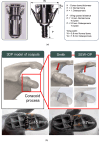
Coracoclavicular (CC) ligament reconstruction model and fixation outcomes. (a) Experimental setup using a traditional hook plate clasped beneath the acromion. (b) Fixation with the SEW anchor implanted into the coracoid process. (c) Radiograph confirming successful deployment of SEW anchor wings after implantation. (d) Hook plate fixation resulting in acromial fracture and structural disintegration. (e) SEW-NP anchors (wings not deployed) showing individual anchor pull-out failure. (f) SEW-OP anchors (wings deployed) resisting individual pull-out and instead failing together with the coracoid bone block, demonstrating superior fixation strength.
Figure 6
Figure 6
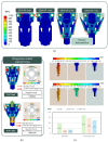
FE analysis of SEW and control anchors under normal and osteoporotic bone conditions. (a) Top: von Mises stress distribution showing concentration around the suture holes and bone–anchor contact areas, with plastic deformation occurring at the wing bottoms. (b) Bottom left: reaction forces at the wing–bone interface under cortical bone thicknesses of H = 2 mm (normal bone, 93.0 N) and H = 1 mm (osteoporotic bone, 148.7 N). (c) Bottom right: displacement patterns of Smith, SEW-NP, and SEW-OP anchors in normal and osteoporotic bone models. SEW-OP exhibited the lowest intra-bone displacement (0.07 mm in normal bone and 0.25 mm in osteoporotic bone), indicating enhanced initial fixation stability compared with Smith and SEW-NP anchors.
Figure 7
Figure 7
SEW anchor design parameters and implantation comparison in the coracoid process model. (a) Dimensional features of the SEW anchor, including cortical bone thickness (H) and wing elevation distance (P). Under normal bone conditions (H = 2 mm), P was approximately 4.5 pitches (3.0 mm), whereas under osteoporotic conditions (H = 1 mm), P increased to 7.5 pitches (1.9 mm). (b) 3D-printed scapula model showing anchor implantation. Compared with the Smith anchor, which required a lateral insertion path and an implantation depth of ~15.8 mm, the SEW-OP anchor was successfully implanted closer to the coracoid base with a reduced depth of ~9.7 mm, demonstrating suitability for limited bone volume.

References

    1. Chaudhry S., Dehne K., Hussain F. A review of suture anchors. Orthop. Trauma. 2019;33:263–270. doi: 10.1016/j.mporth.2016.12.001. - DOI
    1. Braunstein V., Ockert B., Windolf M., Sprecher C.M., Mutschler W., Imhoff A., Postl L.K.L., Biberthaler P., Kirchhoff C. Increasing pullout strength of suture anchors in osteoporotic bone using augmentation—A cadaver study. Clin. Biomech. 2015;30:243–247. doi: 10.1016/j.clinbiomech.2015.02.002. - DOI - PubMed
    1. Horoz L., Hapa O., Barber F.A., Hüsemoğlu B., Özkan M., Havitçioğlu H. Suture anchor fixation in osteoporotic bone: A biomechanical study in an ovine model. Arthrosc. J. Arthrosc. Relat. Surg. 2017;33:68–74. doi: 10.1016/j.arthro.2016.05.040. - DOI - PubMed
    1. Rosso C., Weber T., Dietschy A., de Wild M., Müller S. Three anchor concepts for rotator cuff repair in standardized physiological and osteoporotic bone: A biomechanical study. J. Shoulder Elb. Surg. 2020;29:e52–e59. doi: 10.1016/j.jse.2019.07.032. - DOI - PubMed
    1. Yang Y.S., Shih C.A., Fang C.J., Huang T.T., Hsu K.L., Kuan F.C., Su W.R., Hong C.K. Biomechanical comparison of different suture anchors used in rotator cuff repair surgery-all-suture anchors are equivalent to other suture anchors: A systematic review and network meta-analysis. J. Exp. Orthop. 2023;10:45. doi: 10.1186/s40634-023-00608-w. - DOI - PMC - PubMed

LinkOut - more resources