. 2025 Nov-Dec;22(6):1025-1043.
doi: 10.21873/cgp.20553.

Oxysterol-binding Protein-like 3 Promotes Tumor Progression by Regulating Apoptosis and Angiogenesis in Colorectal Cancer

Affiliations

Oxysterol-binding Protein-like 3 Promotes Tumor Progression by Regulating Apoptosis and Angiogenesis in Colorectal Cancer

Chan-Muk Im et al. Cancer Genomics Proteomics. 2025 Nov-Dec.

Abstract

Background/aim: Oxysterol-binding protein-like 3 (OSBPL3) is a member of the intracellular lipid receptor and transporter protein family involved in regulating lipid metabolism. Altered OSBPL3 expression has been observed in various cancers, where it has been associated with both oncogenic and tumor-suppressive roles. However, its precise functions and underlying mechanisms in colorectal cancer (CRC) remain unclear. This study aimed to investigate the role of OSBPL3 in CRC cells and evaluate its prognostic significance.

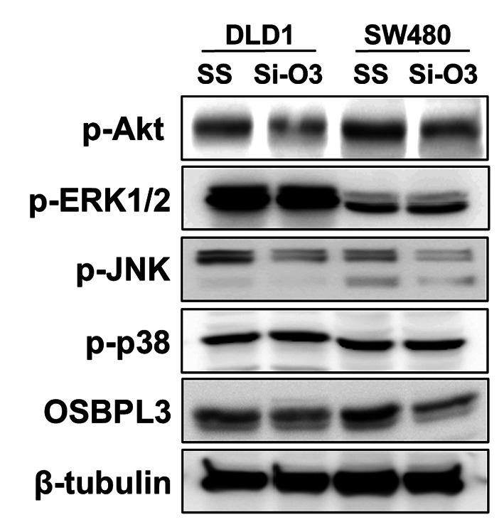
Materials and methods: A small interfering RNA vector targeting OSBPL3 was employed to silence its expression in CRC cell lines. OSBPL3 levels in CRC tissues were assessed using reverse transcription-polymerase chain reaction and immunohistochemistry. Tumor cell apoptosis, proliferation, and angiogenesis were evaluated via a terminal deoxynucleotidyl transferase-mediated dUTP nick-end labeling assay, and immunohistochemical staining for Ki-67 and CD34.

Results: OSBPL3 silencing inhibited tumor cell migration and invasion in CRC. OSBPL3 knockdown reduced proliferation and induced apoptosis through caspase activation and cell cycle arrest mediated by the regulation of cyclins, cyclin dependent kinases (CDKs), and CDK inhibitors. OSBPL3 suppression impaired the invasion and tube formation of human umbilical vein endothelial cells by down-regulating angiogenic factors and up-regulating angiostatic factors. OSBPL3 expression correlated significantly with tumor size, tumor stage, invasion depth, lymph node involvement, distant metastasis, and reduced survival. The apoptotic index was lower and microvessel density was higher in OSBPL3-positive tumors than in OSBPL3-negative tumors.

Conclusion: OSBPL3 contributes to CRC progression by regulating tumor cell apoptosis and angiogenesis.

Keywords: Oxysterol-binding protein-like 3; angiogenesis; apoptosis; colorectal neoplasm; prognosis.


Conflict of interest statement

The Authors declare that they have no conflicts of interest in regard to this study.

Figures

Figure 1
Figure 1
Expression of OSBPL3 in CRC cells. (A) Expression of OSBPL3 protein in human CRC cell lines, including DLD1, HCT116, SW480, Caco-2, HT-29, and SW1116. (B) Expression of OSBPL3 protein following transfection with scramble siRNA or OSBPL3 siRNA in DLD1 and SW480 cells. Western blot analysis confirmed the effective knockdown of OSBPL3 protein by using Si-O3. OSBPL3, Oxysterol-binding protein-like 3; CRC, colorectal cancer; SS, scramble siRNA; Si-O3, OSBPL3 siRNA.
Figure 2
Figure 2
Effect of OSBPL3 knockdown on the proliferation, apoptosis, and cell cycle of CRC cells. (A) Effect of OSBPL3 knockdown on the proliferation of CRC cells. Cell viability, as indicated by absorbance, significantly decreased in Si-O3-transfected DLD1 and SW480 cells on day 3 (mean±SD, n=3; *p<0.05). (B) Effect of OSBPL3 knockdown on the apoptosis of CRC cells. The proportion of apoptotic cells was increased following Si-O3 transfection compared with SS (10.3% vs. 14.8% and 9.8% vs. 15.3% in DLD1 and SW480 cells, respectively). (C) Expression levels of caspases and apoptosis-related proteins. The levels of cleaved caspase-3, -9, and PARP increased after OSBPL3 knockdown in DLD1 and SW480 cells. The expression of the anti-apoptotic protein Bcl-xL decreased. (D) Effect of OSBPL3 knockdown on cell cycle distribution. Knockdown caused the accumulation of DLD1 and SW480 cells in the subG1 phase. Data represent one of three independent experiments. (E) Expression levels of cyclins, CDKs, and CDK inhibitors. OSBPL3 knockdown significantly reduced the cyclin B1 and CDK4 levels and increased the p27 expression in DLD1 and SW480 cells. OSBPL3, Oxysterol-binding protein-like 3; CRC, colorectal cancer; SS, scramble siRNA; Si-O3, OSBPL3 siRNA; SD, standard deviation; PARP, poly (ADP-ribose) polymerase; CC, cleaved caspase; CDK, cyclin-dependent kinase.
Figure 2
Figure 2
Effect of OSBPL3 knockdown on the proliferation, apoptosis, and cell cycle of CRC cells. (A) Effect of OSBPL3 knockdown on the proliferation of CRC cells. Cell viability, as indicated by absorbance, significantly decreased in Si-O3-transfected DLD1 and SW480 cells on day 3 (mean±SD, n=3; *p<0.05). (B) Effect of OSBPL3 knockdown on the apoptosis of CRC cells. The proportion of apoptotic cells was increased following Si-O3 transfection compared with SS (10.3% vs. 14.8% and 9.8% vs. 15.3% in DLD1 and SW480 cells, respectively). (C) Expression levels of caspases and apoptosis-related proteins. The levels of cleaved caspase-3, -9, and PARP increased after OSBPL3 knockdown in DLD1 and SW480 cells. The expression of the anti-apoptotic protein Bcl-xL decreased. (D) Effect of OSBPL3 knockdown on cell cycle distribution. Knockdown caused the accumulation of DLD1 and SW480 cells in the subG1 phase. Data represent one of three independent experiments. (E) Expression levels of cyclins, CDKs, and CDK inhibitors. OSBPL3 knockdown significantly reduced the cyclin B1 and CDK4 levels and increased the p27 expression in DLD1 and SW480 cells. OSBPL3, Oxysterol-binding protein-like 3; CRC, colorectal cancer; SS, scramble siRNA; Si-O3, OSBPL3 siRNA; SD, standard deviation; PARP, poly (ADP-ribose) polymerase; CC, cleaved caspase; CDK, cyclin-dependent kinase.
Figure 3
Figure 3
Effect of OSBPL3 on the migration and invasion of CRC cells. (A) Representative images and quantitative graphs illustrating the effect of OSBPL3 knockdown on CRC cell migration. A wound healing assay was performed using siRNA-transfected cells, and relative healing distances were presented as mean±SD (n=3; *p<0.05). The artificial wound gap in SS-transfected cells became significantly narrower than that in Si-O3-transfected cells at 54 h in DLD1 and SW480 cells. (B) Representative images with corresponding quantitative graphs demonstrating the effect of OSBPL3 knockdown on CRC cell invasion. An invasion assay was conducted using siRNA-transfected cells. Invading cells were stained, counted, and graphed for each group. The number of invading Si-O3-transfected cells was significantly lower than that of SS-transfected DLD1 and SW480 cells (mean±SD, n=3; *p<0.05). (C) Representative images and quantitative data showing the effect of OSBPL3 knockdown on extracellular matrix-related proteins. OSBPL3 knockdown reduced the expression levels of vimentin, MMP-2, MMP-9, snail, and slug and increased the expression of E-cadherin in DLD1 and SW480 cells. OSBPL3, Oxysterol-binding protein-like 3; CRC, colorectal cancer; SS, scramble siRNA; Si-O3, OSBPL3 siRNA; SD, standard deviation; MMP, matrix metalloproteinase.
Figure 3
Figure 3
Effect of OSBPL3 on the migration and invasion of CRC cells. (A) Representative images and quantitative graphs illustrating the effect of OSBPL3 knockdown on CRC cell migration. A wound healing assay was performed using siRNA-transfected cells, and relative healing distances were presented as mean±SD (n=3; *p<0.05). The artificial wound gap in SS-transfected cells became significantly narrower than that in Si-O3-transfected cells at 54 h in DLD1 and SW480 cells. (B) Representative images with corresponding quantitative graphs demonstrating the effect of OSBPL3 knockdown on CRC cell invasion. An invasion assay was conducted using siRNA-transfected cells. Invading cells were stained, counted, and graphed for each group. The number of invading Si-O3-transfected cells was significantly lower than that of SS-transfected DLD1 and SW480 cells (mean±SD, n=3; *p<0.05). (C) Representative images and quantitative data showing the effect of OSBPL3 knockdown on extracellular matrix-related proteins. OSBPL3 knockdown reduced the expression levels of vimentin, MMP-2, MMP-9, snail, and slug and increased the expression of E-cadherin in DLD1 and SW480 cells. OSBPL3, Oxysterol-binding protein-like 3; CRC, colorectal cancer; SS, scramble siRNA; Si-O3, OSBPL3 siRNA; SD, standard deviation; MMP, matrix metalloproteinase.
Figure 4
Figure 4
Effect of OSBPL3 knockdown on the angiogenesis of CRC cells. (A) Representative images and statistical analyses depicting the effect of OSBPL3 knockdown on HUVECs invasion. The invasion of HUVECs cultured in CM from Si-O3-transfected DLD1 and SW480 cells significantly decreased compared to that of HUVECs cultured in CM from SS-transfected cells (p=0.019 and p=0.031, respectively). (B) Representative images alongside quantitative graphs presenting the effect of OSBPL3 knockdown on endothelial tube formation. CM from Si-O3-transfected DLD1 and SW480 cells significantly inhibited endothelial tube formation compared with CM from SS-transfected cells (p=0.043 and p=0.013, respectively). (C) Representative images and quantitative graphs demonstrating the effect of OSBPL3 knockdown on the expression of angiogenesis-related proteins. OSBPL3 knockdown decreased the expression levels of the pro-angiogenic factors HIF-1α, VEGF-A, and VEGF-D and increased the expression of the anti-angiogenic factor angiostatin in DLD1 and SW480 cells. OSBPL3, Oxysterol-binding protein-like 3; CRC, colorectal cancer; SS, scramble siRNA; Si-O3, OSBPL3 siRNA; CM, conditioned media; HUVECs, human umbilical vein endothelial cells; HIF-1α, Hypoxia-inducible factor-1α; VEGF, vascular endothelial growth factor. Each experiment was repeated three times. *p<0.05 vs. control.
Figure 5
Figure 5
Effect of OSBPL3 knockdown on oncogenic signaling pathways involved in the regulation of the OSBPL3 expression in CRC cells. The phosphorylation levels of Akt and JNK were decreased following OSBPL3 knockdown, whereas the phosphorylation levels of ERK1/2 and p38 remained unchanged. OSBPL3, Oxysterol-binding protein-like 3; CRC, colorectal cancer; SS, scramble siRNA; Si-O3, OSBPL3 siRNA; ERK1/2, extracellular signal-regulated kinase1/2; JNK, c-Jun N-terminal kinase.
Figure 6
Figure 6
Expression of OSBPL3 in CRC and metastatic lymph node tissues. (A) Expression images and band quantification graphs of OSBPL3 mRNA levels in colorectal cancer tissue and paired normal mucosa by reverse-transcription PCR. The mRNA expression of OSBPL3 was up-regulated in cancer tissues compared with that in the paired normal mucosa in fresh colonoscopic biopsy specimens. Each bar represents the mean±SD of 20 cases. *p<0.05 vs. normal colorectal mucosa. (B) Immunohistochemical staining of OSBPL3 in normal colorectal mucosa and colorectal cancer tissue. The immunohistochemical staining of OSBPL3 protein showed weak or no staining in normal colorectal mucosa. Conversely, cancer cells exhibited strong cytoplasmic staining, but the tumor stroma had no detectable staining (original magnification, ×100 and ×400). (C) Immunohistochemical staining of OSBPL3 in non-metastatic lymph node and metastatic lymph node (original magnification: ×400). OSBPL3 immunostaining in metastatic lymph nodes was significantly stronger than that in non-metastatic lymph nodes. (D) The overall immunostaining score was significantly higher in metastatic lymph node tissues than in non-metastatic lymph node tissues (*p<0.05; original magnification: ×400). OSBPL3, Oxysterol-binding protein-like 3; CRC, colorectal cancer; T; colorectal cancer tissue, N; paired normal colorectal mucosa, NL; non-metastatic lymph node, ML; metastatic lymph node.
Figure 6
Figure 6
Expression of OSBPL3 in CRC and metastatic lymph node tissues. (A) Expression images and band quantification graphs of OSBPL3 mRNA levels in colorectal cancer tissue and paired normal mucosa by reverse-transcription PCR. The mRNA expression of OSBPL3 was up-regulated in cancer tissues compared with that in the paired normal mucosa in fresh colonoscopic biopsy specimens. Each bar represents the mean±SD of 20 cases. *p<0.05 vs. normal colorectal mucosa. (B) Immunohistochemical staining of OSBPL3 in normal colorectal mucosa and colorectal cancer tissue. The immunohistochemical staining of OSBPL3 protein showed weak or no staining in normal colorectal mucosa. Conversely, cancer cells exhibited strong cytoplasmic staining, but the tumor stroma had no detectable staining (original magnification, ×100 and ×400). (C) Immunohistochemical staining of OSBPL3 in non-metastatic lymph node and metastatic lymph node (original magnification: ×400). OSBPL3 immunostaining in metastatic lymph nodes was significantly stronger than that in non-metastatic lymph nodes. (D) The overall immunostaining score was significantly higher in metastatic lymph node tissues than in non-metastatic lymph node tissues (*p<0.05; original magnification: ×400). OSBPL3, Oxysterol-binding protein-like 3; CRC, colorectal cancer; T; colorectal cancer tissue, N; paired normal colorectal mucosa, NL; non-metastatic lymph node, ML; metastatic lymph node.
Figure 7
Figure 7
Kaplan-Meier survival curve correlating overall survival with the negative (solid line) and positive expression (dotted line) of OSBPL3. OSBPL3, Oxysterol-binding protein-like 3.
Figure 8
Figure 8
Assessment of tumor cell apoptosis, proliferation, and angiogenesis in CRCs. (A) Detection of apoptotic cells and bodies by TUNEL staining. (B) Square box in A. Apoptotic bodies were identified by the presence of pyknotic nuclei surrounded by a condensed cytoplasm and separated from neighboring cells by a clear halo. (C) Immunohistochemical staining of Ki-67 in CRC tissue. (D) Square box in C. Ki-67 immunoreactivity was observed in the nuclei of proliferating cancer cells. (E) Immunohistochemical staining of CD34 in CRC tissue. (F) Square box in D. Antibody-specific vessels were defined as single brown-stained cells or clusters of endothelial cells clearly distinguishable from adjacent microvessels and tumor cells (scale bar corresponds to 10 μm). OSBPL3, Oxysterol-binding protein-like 3; CRC, colorectal cancer; TUNEL, terminal deoxynucleotidyl transferase (TdT)-mediated deoxyuridine triphosphate (dUTP) nick end labeling.

References

    1. Mattiuzzi C, Sanchis-Gomar F, Lippi G. Concise update on colorectal cancer epidemiology. Ann Transl Med. 2019;7(21):609. doi: 10.21037/atm.2019.07.91. - DOI - PMC - PubMed
    1. Hossain MS, Karuniawati H, Jairoun AA, Urbi Z, Ooi J, John A, Lim YC, Kibria KMK, Mohiuddin AKM, Ming LC, Goh KW, Hadi MA. Colorectal cancer: a review of carcinogenesis, global epidemiology, current challenges, risk factors, preventive and treatment strategies. Cancers (Basel) 2022;14(7):1732. doi: 10.3390/cancers14071732. - DOI - PMC - PubMed
    1. Hančinová M, Ondruš D, Ondrušová M, Mriňáková B. Colorectal carcinoma - epidemiology, risk factors, prognostic biomarkers. Klin Onkol. 2025;38(1):16–24. doi: 10.48095/ccko202516. - DOI - PubMed
    1. Anbari K, Ghanadi K. Colorectal cancer: risk factors, novel approaches in molecular screening and treatment. Int J Mol Cell Med. 2025;14(1):576–605. doi: 10.22088/IJMCM.BUMS.14.1.576. - DOI - PMC - PubMed
    1. Alampritis G, Thoukididou SN, Ramos M, Georgiou P, Kalofonou M, Simillis C. Diagnostic value of genetic and epigenetic biomarker panels for colorectal cancer detection: a systematic review. Int J Colorectal Dis. 2025;40(1):125. doi: 10.1007/s00384-025-04904-y. - DOI - PMC - PubMed

MeSH terms

Substances

LinkOut - more resources