. 2025 Oct 28;15(1):37704.
doi: 10.1038/s41598-025-21534-0.

Tauroursodeoxycholic acid-induced increase in ectopic muscle mineralization occurs exclusively in dystrophic muscles and is independent of endoplasmic reticulum stress

Affiliations

Tauroursodeoxycholic acid-induced increase in ectopic muscle mineralization occurs exclusively in dystrophic muscles and is independent of endoplasmic reticulum stress

Justyna Róg et al. Sci Rep. .

Abstract

Calcification of dystrophic skeletal muscles was described previously and attributed, among others, to ER-stress, elevated phosphate concentration and chronic inflammation. Tauroursodeoxycholic acid (TUDCA) is considered an artificial chaperone protecting cells against ER-stress thus could prevent an ectopic mineralisation of soft tissues. Because an enhanced ER-stress is a feature of dystrophic muscles and it promotes soft tissue mineralisation we hypothesised that TUDCA treatment should reduce mineral deposits in dystrophic skeletal muscles, and tested this concept using two mouse models of DMD. Four-week old mdx, mdxβetageo and w/t mice were administered TUDCA in drinking water for 4 weeks. At 8 weeks, following tissue-clearing and calcium minerals staining with alizarin, mineralisation was evaluated using whole body scanning. Additionally, isolated skeletal muscles were analysed by Western blotting for ER-stress and calcification markers, and using various microscopic methods. Enzymatic activity of alkaline phosphatase was also assayed. Unexpectedly, TUDCA enhanced calcification of dystrophic but not dystrophin-positive muscles. TUDCA did not affect the elevated ER-stress markers found in dystrophic muscles nor impact pro-calcifying proteins RUNX2, Osterix and BMP2/4, which were also overexpressed in dystrophic muscles. The alkaline phosphatase levels, which were reduced in dystrophic muscles, were not affected by this treatment. The increase in ectopic calcification in dystrophic muscles induced by TUDCA is specific to muscles lacking dystrophin. This effect is not linked to the alleviation of ER stress or the overexpression of proteins directly involved in calcium mineral accumulation.


Conflict of interest statement

Declarations. Competing interests: The authors declare no competing interests. Ethics approval: Every effort was made to minimize the number of animals used and the level of stress they endure. All methods we used including anaesthesia and euthanasia were consistent with the commonly accepted norms of veterinary best practice. The study was carried out in accordance with relevant guidelines and regulations as described by the ARRIVE guidelines (PLoS Bio 8(6), e1000412, 2010). The experimental procedures also complied with the Polish Law on Experiments on Animals, which implements the European Council Directive, and were approved by the 1st Local Ethical Committee for Animal Experimentation in Warsaw (Permit Number: 1279/2022).

Figures
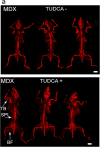
Fig. 1
Fig. 1
Whole-body analysis of ectopic calcifications in mdx treated with TUDCA. Tissue clearing and Alizarin red staining show distribution of ectopic calcification across the entire musculature. (a) Representative epifluorescent images reveal sites of myofiber calcification. (b) Three-dimensional light-sheet microscopy data of selected sites of ectopic calcification. (c) Percentage mineralization and cumulative frequency distributions in triceps pars brachii, biceps pars femoris and spinalis pars lumborum). Unpaired t-test and two-sample Kolmogorov–Smirnov test were performed. Data are expressed as means ± SD for three animals (independent biological replicates). *P < 0.05.
Fig. 1
Fig. 1
Whole-body analysis of ectopic calcifications in mdx treated with TUDCA. Tissue clearing and Alizarin red staining show distribution of ectopic calcification across the entire musculature. (a) Representative epifluorescent images reveal sites of myofiber calcification. (b) Three-dimensional light-sheet microscopy data of selected sites of ectopic calcification. (c) Percentage mineralization and cumulative frequency distributions in triceps pars brachii, biceps pars femoris and spinalis pars lumborum). Unpaired t-test and two-sample Kolmogorov–Smirnov test were performed. Data are expressed as means ± SD for three animals (independent biological replicates). *P < 0.05.
Fig. 2
Fig. 2
Three-dimensional muscle analysis of ectopic calcifications. Triceps brachii from macroscopically prescreened cleared mice was isolated and imaged in light-sheet fluorescence microscopy for detailed comparison. Column in green presents Alizarin red signal along with autofluorescence of the entire muscle, column in red—segmented Alirazin red signal, n = 6 muscles per group. In the bottom Triceps brachii muscles isolated from w/t animals, both treated and untreated with TUDCA, do not present any calcifications. Scale bar = 1 mm.
Fig. 3
Fig. 3
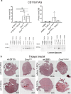
Effects of TUDCA on calcification and inflammation in Dmdmdx and Dmdmdx-βgeo triceps muscle. (a) Western blot analysis of CD11b protein expression in wild-type and dystrophic triceps. Graphs represent data from three mice and corresponding western blots are shown below. (b) Mineralization in C57BL10 and Dmdmdx (left), BL6 and Dmdmdx-βgeo (right). Calcium deposits were detected by Alizarin Red S and are visible as red clumps visualized under the light microscope (10 × magnification). (c) Confocal images of CD11b immunolocalization. The nuclei were labelled using Hoechst (blue colour). Tissues shown in panels b and c are adjacent sections from the same muscle, which demonstrates the co-existence of mineral deposits and CD11b stained macrophages in the same area.
Fig. 3
Fig. 3
Effects of TUDCA on calcification and inflammation in Dmdmdx and Dmdmdx-βgeo triceps muscle. (a) Western blot analysis of CD11b protein expression in wild-type and dystrophic triceps. Graphs represent data from three mice and corresponding western blots are shown below. (b) Mineralization in C57BL10 and Dmdmdx (left), BL6 and Dmdmdx-βgeo (right). Calcium deposits were detected by Alizarin Red S and are visible as red clumps visualized under the light microscope (10 × magnification). (c) Confocal images of CD11b immunolocalization. The nuclei were labelled using Hoechst (blue colour). Tissues shown in panels b and c are adjacent sections from the same muscle, which demonstrates the co-existence of mineral deposits and CD11b stained macrophages in the same area.
Fig. 4
Fig. 4
Effect of TUDCA on ER-stress marker levels in lysates from Dmdmdx, Dmdmdx-βgeo and wild-type mouse triceps muscle. Data show mean values ± S.D. For three independent experiments. Statistical significance at p < 0.05 is indicated.
Fig. 5
Fig. 5
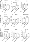
Effect of TUDCA on unfolded protein response marker levels in lysates from Dmdmdx and wild-type mouse triceps. (a, b, c) Indicate three distinct UPR pathways. Data show mean values ± S.D. for three individual animals. Statistical significance at p < 0.05 is indicated. Corresponding western blots are shown in Suppl. CHOP—C/EBP homologous protein, eIFalpha—Eukaryotic Initiation Factor 2, IRE alpha—inositol-requiring enzyme 1α, XBP1-X-box binding protein 1, ATF4 -Activating transcription factor 4.
Fig. 6
Fig. 6
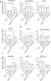
Effect of TUDCA on unfolded protein response marker levels in lysates from Dmdmdx-βgeo and wild-type mouse triceps. (a, b, c) Indicate three distinct UPR pathways. Data show mean values ± S.D. for three individual animals. Statistical significance at p < 0.05 is indicated. Corresponding western blots are shown in Suppl. 2 and 3. Abbreviations as in Fig. 5.
Fig. 7
Fig. 7
Expression of osteogenic factors Osterix, RUNX2, BMP2 and TNAP in dystrophic muscles. Graphs present relative protein levels in lysates from triceps muscle isolated from w/t and dystrophic mice. Below the relevant Western blots are shown. Upper Row: Dmdmdx and C57Bl10 samples. Lower row: Dmdmdx-βgeo and C57Bl6 samples. Graphs show mean values ± S.D. for three independent animals. Statistcal significance is indicated. Corresponding western blots are shown in Suppl. 2 and 3.
Fig. 7
Fig. 7
Expression of osteogenic factors Osterix, RUNX2, BMP2 and TNAP in dystrophic muscles. Graphs present relative protein levels in lysates from triceps muscle isolated from w/t and dystrophic mice. Below the relevant Western blots are shown. Upper Row: Dmdmdx and C57Bl10 samples. Lower row: Dmdmdx-βgeo and C57Bl6 samples. Graphs show mean values ± S.D. for three independent animals. Statistcal significance is indicated. Corresponding western blots are shown in Suppl. 2 and 3.
Fig. 8
Fig. 8
Effect of TUDCA on alkaline phosphatase activity. Enzymatic activity was measured in homogenates prepared from the Dmdmdx peritoneal macrophages isolated from control (non-treated) mice and Dmdmdx-βgeo triceps isolated from control or TUDCA-treated mice. To further asses a putative effect of TUDCA on TNAP activity an assay was performed where 100 μM TUDCA was added directly before measurement. Collected data from three individual animals (biological replicates).

References

    1. Yiu, E. M. & Kornberg, A. J. Duchenne muscular dystrophy. J. Paediatr. Child Health51(8), 759–764. 10.1111/jpc.12868 (2015). - PubMed
    1. Batti Angulski, A. et al. Duchenne muscular dystrophy: Disease mechanism and therapeutic strategies. Front. Physiol.26(14), 1183101. 10.3389/fphys.2023.1183101 (2023). - PMC - PubMed
    1. Snow, W. M., Anderson, J. E. & Jakobson, L. S. Neuropsychological and neurobehavioral functioning in Duchenne muscular dystrophy: a review. Neurosci Biobehav Rev.37(5), 743–752. 10.1016/j.neubiorev.2013.03.016 (2013). - PubMed
    1. Rufo, A. et al. Mechanisms inducing low bone density in Duchenne muscular dystrophy in mice and humans. J. Bone Miner. Res.26(8), 1891–1903. 10.1002/jbmr.410 (2011). - PMC - PubMed
    1. Tsaknakis, K. et al. Reduced bone mineral density in adolescents with Duchenne Muscular Dystrophy (DMD) and scoliosis. Osteoporos Int.33(9), 2011–2018. 10.1007/s00198-022-06416-9 (2022). - PMC - PubMed

MeSH terms

Substances

LinkOut - more resources