. 2025 Oct 21;15(20):2660.
doi: 10.3390/diagnostics15202660.

A Revised Concept for Ocular Surface Imprinting: Easy-to-Use Device for Morphological and Biomolecular-Based Differential Diagnosis

Affiliations

A Revised Concept for Ocular Surface Imprinting: Easy-to-Use Device for Morphological and Biomolecular-Based Differential Diagnosis

Bijorn Omar Balzamino et al. Diagnostics (Basel). .

Abstract

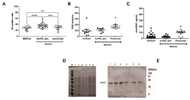
Background/objectives: The continuous necessity to support biostrumental data with biolomecular data collected using non-invasive tools is influencing the world of ocular surface devices. The ocular imprint still represents a non-invasive and safety technique for collecting corneal and conjunctival epithelia in an easy way, as performed in human and veterinary clinics. Although used in clinical practice since 1977, operators might benefit from improvements in these techniques, especially in terms of handling and management. Methods: Herein, by reporting the design and characteristics of a patent of ocular surface sampling (the SurfAL pen and periocular-assisted SurfAL pen; PCT WO2016IB51474 20160316), we performed a validation and analysis of its value compared to gold standards. The level-headedness and advantages of this device were verified in 15 sclerocorneal specimens (sampling advantages) and tested in 25 volunteers (handling and operator efficiency, as well as frequency of discomfort in volunteers). Morphological as well as biomolecular analyses were used to compare SurfAL devices with conventional ones. Results: The easy management of SurfAL pens and the good detection of epithelial/goblet cells were confirmed. The SurfAL pen was found to be smart and suitable for routine analysis, as confirmed by quick and reproducible onsite sampling. Periocular-assisted SurfAL pen was comparable in terms of sampling quality but less comparable in terms of subject confidence due to its geometry. Conclusions: This study suggests that the SurfAL pen and periocular-assisted SurfAL pen might represent an additional and hands-on way of sampling ocular surface cells and improve the diagnostic route in ophthalmology.

Keywords: clinical practice; epithelial imprints; goblet cells; ocular surface; point-of-care device; sampling device.


Conflict of interest statement

The authors declare that there is no conflict of interest.

Figures

Figure 1
Figure 1
Description of SurfAL pen. (A) Front view of the hollow for the insertion of the MilliCell membrane support or the single membrane. Dimensions are summarized in the box. (B,C) Pick-up pen: stick and dimensions. As shown in A, the SurfAL pen has a full cylindrical shape (17 mm outer diameter). The 14.5 mm inner diameter can hold the nitrocellulose membrane (Millicell, Millipore). The total length is approximately 87 mm, and the weight is about 10 g, allowing a perfect grip for the operator’s hand. Mounting geometry has been designed to be easy to handle for operators. Depending on packaging, the device can be disposable or refillable with the commercial membrane device.
Figure 2
Figure 2
(A) The periocular-assisted SurfAL pen device (combo device) has a cylindrical geometry with a disk moving on four linear rails containing specific springs. The device controls the imprinting force on the ocular surface. The round shape (50 mm outside and 40 mm inside) provides a better adherence to the ocular orbit and a complete wrapping of the eye orbit in a comfortable condition for the patient. The eyelid motion is partially blocked, and the device does not cause any sense of discomfort or constriction to the patient. The height is approximately 30 mm, and the weight is about 50 g, excluding the inner springs. (B) Upper view of the disk with a hole for pick-up stick insertion. Related measurements are reported. The mobile disk has one hole of 17 mm to allow the insertion of the SurfAL pen, as highlighted in green, allowing sampling at four different quadrants on the eye surface. To compensate for the curvature of the eye, avoiding incorrect inclinations of the SurfAL pen, the hole is inclined at an angle of 10° to the center of the disk.
Figure 3
Figure 3
Sampling procedures. (A) A particular corneoscleral rim in the holder, as backlit by optic fibers. (B) a time-lapse of the sampling procedure. Both time, pressure and inclination were reproduced as in Clinique.
Figure 4
Figure 4
A radar chart showing the results of the analysis of questionnaires. This spider chart represents the allocated qualities of the two separated subsystems and the use of the combo device. The radar chart shows the differences in performance metrics of subsystem A (A), subsystem B (B) and the combo subsystem A + B (C).
Figure 5
Figure 5
Sampling procedures. From left to right are the following procedures(A,C): (A) Millicell support; (B) SurfAL pen; (C) periocular-assisted SurfAL device (combo SurfAL). The volunteer adhered to illustrative purposes.
Figure 6
Figure 6
Microscopy validation. Imprints from the three different methods of sampling were stained for basal cytology (PAS staining) or immunofluorescent analysis of mucins (muc5AC) and cytokeratins (CK12; CK19). PAS-positive magenta-stained goblet cells in the upper panels (AC); CK12/19-immunoreactive (DF) and Muc5AC-positive cells (F,G) are shown, respectively, in the middle and lower panels. The number of PAS-positive or mucin-immunoreactive cells represents an index of the healthiness of the ocular surface and is therefore used for diagnostic purposes. Rows included as follows: (A,D,G), MilliCell; (B,E,H), SurfAL pen; (C,F,I), periocular-assisted SurfAL pen device.
Figure 7
Figure 7
Microscopic vs. biochemical validations on imprints from the three different methods of sampling. (A) Cell counting of Acidic Phosphatases immunolabeled cells (A) and IntDen analysis (MFI) for PAS (B) and muc5AC (C) positive cells per optic field (×20), respectively, in membranes from Millicell, SurfAL pen, and periocular device. (D) Respectively 4–20% SDS-PAGE and (E) cytokeratin (CK) 19 probed immunoblot from imprints: Millicell (1,2), SurfAL pen (3,4), and periocular (5,6) devices. MFI, Mean Fluorescent Intensity; M, sizer; L, ladder. Significant changes are pointed by asterisks (ns not significant; *** p < 0.005; **** p < 0.001 REST–ANOVA–Tukey–Kramer post hoc).
Figure 8
Figure 8
Molecular validations. Total RNA extraction and transcript amplification were carried out on imprints from Millicell and SurfAL devices. The panel shows the following: (A) total RNA expression in comparison between Millicell and SurfAL; (B) representative amplicons (muc5ACmRNA) in membranes from all devices run in duplicate (from left to right: size marker (L), Millicell (1,2), SurfAL pen (3,4) and periocular (5,6) devices); and (C) scatter-plots showing the Ct values for GAPDH (C), HLADR (D), ICAM1 (E), p65NFkB (G), muc5AC (F), and IL6 (H), all Millicell vs. SurfAL pen. Note that the Cts values were used for comparison. Cts and transcript expression values are inversely related. (ns not significant and * p < 0.05 REST–ANOVA–Tukey–Kramer post hoc).
Figure 9
Figure 9
Schematic representation of SurfAL modules and applications. The SurfAL device was designed and developed to assist clinical human and veterinary practice with different diagnostic purposes. (A) SurfAL is a flexible platform to be used for identifying biomarkers representative of different stages of discomfort or disease whenever validated in other studies. The periocular-assisted SurfAL device is shown and allows a more flexible, easy, and precise monolayer sampling for human and veterinary practices. SurfAL loads appropriate membranes (nitrocellulose) as Millicell does. (B) The major points in the procedure of sampling are summarized, highlighting the easy procedure and flexibility of this point-of-care. In private clinical centers as well as in clinical and surgical departments, the point-of-care device might provide quick sampling and delivery to the laboratory for analysis or just an on-site analysis. (C) A list of potential targets suitable for this device, clustered for functional and morphological purposes, is shown. Biomarkers include proteins and peptides, DNA, RNA, and microRNA, as well as infections and microbiome targets, cortisol and neuropeptides, hormones, stress-related proteins, and Reactive Oxygen Species (ROS). Targets can be detected at both biomolecular and morphological levels. A simultaneous detection of biomarkers can be suitable (RNA/DNA/protein), and the morphological analyses represent an additional point for point-of-care applications.

References

    1. Cutrupi F., De Luca A., Di Zazzo A., Micera A., Coassin M., Bonini S. Real Life Impact of Dry Eye Disease. Semin. Ophthalmol. 2023;38:690–702. doi: 10.1080/08820538.2023.2204931. - DOI - PubMed
    1. Stern M.E., Pflugfelder S.C. Inflammation in dry eye. Ocul. Surf. 2004;2:124–130. doi: 10.1016/S1542-0124(12)70148-9. - DOI - PubMed
    1. Kanski J.J. Systemic diseases and the eye: Signs and differential diagnoses. CV Mosby, London, 2001. pp. 241. $79.95. Am. J. Ophthalmol. 2002;133:592.
    1. Aronni S., Cortes M., Sacchetti M., Lambiase A., Micera A., Sgrulletta R., Bonini S. Upregulation of ICAM-1 expression in the conjunctiva of patients with chronic graft-versus-host disease. Eur. J. Ophthalmol. 2006;16:17–23. doi: 10.1177/112067210601600104. - DOI - PubMed
    1. Pisella P.J., Brignole F., Debbasch C., Lozato P.A., Creuzot-Garcher C., Bara J., Saiag P., Warnet J.M., Baudouin C. Flow cytometric analysis of conjunctival epithelium in ocular rosacea and keratoconjunctivitis sicca. Ophthalmology. 2000;107:1841–1849. doi: 10.1016/S0161-6420(00)00347-X. - DOI - PubMed

LinkOut - more resources