. 2025 Sep 30;14(9):5490-5503.
doi: 10.21037/tcr-2025-480. Epub 2025 Sep 22.

CMTM4 overexpression inhibited tumor growth through regulating the function of myeloid-derived suppressor cells in the cervical cancer microenvironment

Affiliations

CMTM4 overexpression inhibited tumor growth through regulating the function of myeloid-derived suppressor cells in the cervical cancer microenvironment

Haiyan Wang et al. Transl Cancer Res. .

Abstract

Background: CMTM4 is involved in various cellular processes, such as cancer progression and immune regulation. However, its precise role in cervical cancer remains unclear. This study aimed to investigate the impact of CMTM4 on HeLa cell behavior, tumorigenesis, myeloid-derived suppressor cell (MDSC) generation, and immunosuppressive effects in the context of cervical cancer.

Methods: HeLa cells were genetically engineered to overexpress CMTM4 or to achieve CMTM4 knockdown. Subsequently, both in vitro and in vivo experiments were performed to systematically evaluate cell proliferation, apoptosis, tumorigenesis, as well as the behavior of immune cells. MDSCs were isolated and treated to analyze the effects of CMTM4 on their function.

Results: Immunohistochemical and Western blot analyses indicated a diminished expression of the CMTM4 protein in cervical cancer tissues relative to adjacent non-cancerous tissues. In vitro experiments revealed that silencing CMTM4 enhanced the viability of HeLa cells and reduced apoptosis, whereas its overexpression produced the opposite effects. Furthermore, CMTM4 overexpression mitigated the immunosuppressive effects of MDSCs. However, the introduction of exogenous MDSCs counteracted the inhibitory impact of CMTM4 on cervical cancer cell proliferation. Mechanistically, CMTM4 knockdown resulted in increased phosphorylation levels of AKT, STAT3, and ERK, while its overexpression led to decreased phosphorylation of these proteins (p-AKT, p-STAT3, and p-ERK). In vivo experiments demonstrated that CMTM4 overexpression significantly reduced tumor volume and weight, downregulated the expression of proliferation markers such as PCNA and Ki67, and inhibited the activation of the AKT/STAT3/ERK signaling pathway. Additionally, CMTM4 overexpression was associated with a reduction in MDSC infiltration within cervical tumor tissues.

Conclusions: CMTM4 emerges as a critical regulator in cervical cancer, influencing both tumor cell behavior and the immune microenvironment.

Keywords: AKT/STAT3/ERK signaling pathway; CMTM4; Cervical cancer; myeloid-derived suppressor cells (MDSCs).


Conflict of interest statement

Conflicts of Interest: All authors have completed the ICMJE uniform disclosure form (available at https://tcr.amegroups.com/article/view/10.21037/tcr-2025-480/coif). The authors have no conflicts of interest to declare.

Figures

Figure 1
Figure 1
The expression of CMTM4 was down-regulated in cervical cancer tissues. (A,B) IHC stain of CMTM4 in the paracancer tissue and carcinoma tissue of cervical cancer patients (×400 magnification). (C,D) CMTM4 expression in carcinoma and paracancer tissues was assessed by western blot assay. β-actin as the internal reference. **, P<0.01 vs. paracancer tissues. DAB, 3,3'-diaminobenzidine; IHC, immunohistochemical.
Figure 2
Figure 2
CMTM4 regulated HeLa cell progression. (A) The effect of CMTM4 on the growth of HeLa cells was detected by CCK-8 test. (B,C) Cell apoptosis was checked by flow cytometry. (D,E) Cell cycle status was assessed by flow cytometry. (F,G) Western blot was used to assay determined Bax, Bcl-2, and cleaved caspase-9 expression. (H,I) Western blot was used to assay determined AKT, STAT3, ERK, p-AKT, p-STAT3, p-ERK expression. *, P<0.05; **, P<0.01 vs. NC siRNA. #, P<0.05; ##, P<0.01 vs. pcDNA-NC. CCK-8, Cell Counting Kit-8; NC, normal control; OD, optical density.
Figure 3
Figure 3
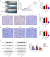
CMTM4 overexpression inhibited HeLa tumorigenesis and decreased AKT/STAT3/ERK signaling pathway in vivo. (A,B) Compared with the AD-NC group, AD-CMTM4 inhibited the tumor volume. (C) Tumor weight were recorded. (D,E) The expression of PCNA was measured using immunohistochemical stain. Magnification is 400×. (F,G) IHC stain of Ki67, magnification is 400×. **P<0.01 vs. AD-NC. (H,I) Western blot was used to assay determined AKT, STAT3, ERK, p-AKT, p-STAT3, p-ERK expression. **, P<0.01 vs. Blank (skin tissue). #, P<0.01 vs. AD-NC. AD, adenovirus; DAB, 3,3'-diaminobenzidine; NC, normal control.
Figure 4
Figure 4
CMTM4 overexpression reduced MDSC infiltration in mouse cervical cancer tumors and in vitro. (A,B) The proportion of MDSCs in spleen, blood and tumor tissues of tumor-bearing mice was determined by flow cytometry. (C,D) The CD11b+/Gr1+ cells in tumor tissues were detected by IHC stain. Magnification is 400×. *, P<0.05; **, P<0.01 vs. AD-NC. (E,F) MDSCs ratio by flow cytometry (CD11b+/Gr1+) in vitro. **, P<0.01 vs. NC siRNA. ##, P<0.01 vs. pcDNA-NC. AD, adenovirus; DAB, 3,3'-diaminobenzidine; IHC, immunohistochemical; MDSC, myeloid-derived suppressor cell; NC, normal control.
Figure 5
Figure 5
CMTM4 overexpression blocked the immunosuppressive effects of MDCSs. (A,B) Cell apoptosis was observed by flow cytometry. (C-E) ELISA is performed to detect secreted flow inflammatory cytokines (IFN-γ, IL-10, and TNF-β) in cell culture supernatants. **, P<0.01 vs. Control. ##, P<0.01 vs. t-MDSCsNC. (F,G) The percentage of proliferating CD3+ T cells in tumor tissues was determined by flow cytometric analysis. (H,I) The proliferation rate of CD3+ T cells in spleen was analyzed by flow cytometry. (J,K) Tumor expression of CD3, was determined by IHC analysis. Magnification is 400×. **, P<0.01 vs. AD-NC. AD, adenovirus; DAB, 3,3'-diaminobenzidine; ELISA, enzyme-linked immunosorbent assay; IHC, immunohistochemical; IL-10, interleukin-10; IFN-γ, interferon-gamma; MDSCs, myeloid-derived suppressor cells; NC, normal control; TNF-β, tumor necrosis factor-beta; WT, wild type.
Figure 6
Figure 6
Exogenous MDSCs reversed the inhibitory effect of CMTM4 on the growth of cervical cancer. (A-C) Tumor volume and tumor weight measurements were conducted. (D,E) Immunohistochemical staining was utilized to assess the expression of PCNA. The magnification used was 400×. (F,G) Ki67 immunohistochemical staining was performed at a magnification of 400×. *, P<0.05; **, P<0.01 vs. AD-NC. #, P<0.05; ##, P<0.01 vs. AD-CMTM4. AD, adenovirus; DAB, 3,3'-diaminobenzidine; MDSC, myeloid-derived suppressor cell; NC, normal control.

References

    1. Abu-Rustum NR, Yashar CM, Arend R, et al. NCCN Guidelines® Insights: Cervical Cancer, Version 1.2024. J Natl Compr Canc Netw 2023;21:1224-33. 10.6004/jnccn.2023.0062 - DOI - PubMed
    1. Perkins RB, Wentzensen N, Guido RS, et al. Cervical Cancer Screening: A Review. JAMA 2023;330:547-58. 10.1001/jama.2023.13174 - DOI - PubMed
    1. Sung H, Ferlay J, Siegel RL, et al. Global Cancer Statistics 2020: GLOBOCAN Estimates of Incidence and Mortality Worldwide for 36 Cancers in 185 Countries. CA Cancer J Clin 2021;71:209-49. 10.3322/caac.21660 - DOI - PubMed
    1. Voelker RA. Cervical Cancer Screening. JAMA 2023;330:2030. 10.1001/jama.2023.21987 - DOI - PubMed
    1. Burmeister CA, Khan SF, Schäfer G, et al. Cervical cancer therapies: Current challenges and future perspectives. Tumour Virus Res 2022;13:200238. 10.1016/j.tvr.2022.200238 - DOI - PMC - PubMed

LinkOut - more resources