. 2025 Oct 24:3:IMAG.a.960.
doi: 10.1162/IMAG.a.960. eCollection 2025.

GOUHFI: A novel contrast- and resolution-agnostic segmentation tool for ultra-high-field MRI

Affiliations

GOUHFI: A novel contrast- and resolution-agnostic segmentation tool for ultra-high-field MRI

Marc-Antoine Fortin et al. Imaging Neurosci (Camb). .

Abstract

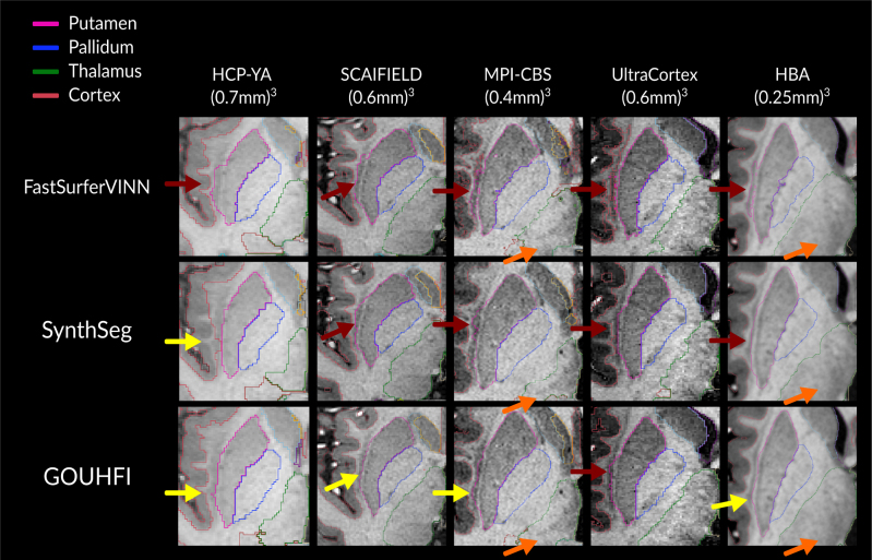
Recently, ultra-high-field MRI (UHF-MRI) has become more available and one of the best tools to study the brain for neuroscientists. One common step in quantitative neuroimaging is to segment the brain into several regions, which has been done using software packages such as FreeSurfer, FastSurferVINN, or SynthSeg. However, the differences between UHF-MRI and 1.5T or 3T images are such that the automatic segmentation techniques optimized at these field strengths usually produce unsatisfactory segmentation results for UHF images. Thus, it has been particularly challenging to perform region-based quantitative analyses as typically done with 1.5-3T data, considerably limiting the potential of UHF-MRI until now. Ultimately, this underscores the crucial need for developing new automatic segmentation techniques designed to handle UHF images. Hence, we propose a novel Deep Learning (DL)-based segmentation technique called GOUHFI: Generalized and Optimized segmentation tool for ultra-high-field images, designed to segment UHF images of various contrasts and resolutions. For training, we used a total of 206 label maps from four datasets acquired at 3T, 7T, and 9.4T. In contrast to most DL strategies, we used a previously proposed domain randomization approach, where synthetic images generated from the 206 label maps were used for training a 3D U-Net. This approach enables the DL model to become contrast agnostic. GOUHFI was tested on seven different datasets and compared with existing techniques such as FastSurferVINN, SynthSeg, and CEREBRUM-7T. GOUHFI was able to segment the six contrasts and seven resolutions tested at 3T, 7T, and 9.4T. Average Dice-Sørensen Similarity Coefficient (DSC) scores of 0.90, 0.90, and 0.93 were computed against the ground truth segmentations at 3T, 7T, and 9.4T, respectively. These results demonstrated GOUHFI's superior performance to competing approaches at each resolution and contrast level tested. Moreover, GOUHFI demonstrated impressive resistance to the typical inhomogeneities observed at UHF-MRI, making it a new powerful segmentation tool allowing the usual quantitative analysis pipelines performed at lower fields to be applied also at UHF. Ultimately, GOUHFI is a promising new segmentation tool, being the first of its kind proposing a contrast- and resolution-agnostic alternative for UHF-MRI without requiring fine tuning or retraining, making it the forthcoming alternative for neuroscientists working with UHF-MRI or even lower field strengths.

Keywords: UHF-MRI; brain segmentation; contrast and resolution agnosticity; deep learning; domain randomization; neuroimaging.


Conflict of interest statement

The authors do not declare any competing interests.

Figures

Fig. 1.
Fig. 1.
Pipeline used to create the training data for GOUHFI. To produce the training data, the sub-millimeter T1w image was used as input to FastSurferVINN in order to create a label map of the whole brain with 35 labels. A new label map was then created by modifying the FastSurferVINN output by adding an extra-cerebral label based on the morphologically modified brain mask (dark gray area surrounding the cerebral cortex on the third sub-figure). This new label map was then used as input to the modified generative model from SynthSeg to create the randomly deformed version of it. Then, the augmented label map was used to generate the synthetic image where, as explained in Billot et al. (2023), a mean and standard deviation are randomly sampled from a normal distribution to generate a noisy signal for each label iteratively. Ultimately, the extra-cerebral label was kept for generating the synthetic images but excluded from the final label map in order to simulate signal surrounding the cortex such as CSF or remaining non-brain tissues from low-quality skull stripping. Finally, the generated synthetic image with its corresponding deformed label map (from which it was generated) was used as the training data (green box).
Fig. 2.
Fig. 2.
Segmentations produced by FastSurferVINN (second column), SynthSeg (third column), and GOUHFI (right column) for one subject in all anatomical planes for the T1w (top) and T2w (bottom) contrasts from the HCP dataset (3T). No segmentations are shown for the T2w image for FastSurferVINN since it only segments T1w images. Dark blue arrows represent regions of discrepancies for the thalamus region with the ground truth for SynthSeg. Turquoise arrows point to cortical regions with limited delineation by SynthSeg. Green arrows show systematic errors with SynthSeg including the claustrum in the putamen label. Yellow arrows show differences in cerebellum WM segmentation. The labels shown and their colors correspond to the FreeSurfer lookup table.
Fig. 3.
Fig. 3.
Segmentations produced by FastSurferVINN (first row), SynthSeg (second row), and GOUHFI (bottom row) for one subject in the coronal (top part) and sagittal (bottom part) planes for the T1w MPRAGE (left), MPM-MTw (second from the left), MPM-PDw (second from the right), and MPM-T1w (right) contrasts from the SCAIFIELD dataset (7T). All images and segmentations have a resolution of (0.6 mm)3. The MPRAGE images have been N4-corrected whereas all MPM contrasts have not. Segmentations from FastSurferVINN are shown for the T1w image only since it only segments T1w images. Blue arrows represent regions of mislabeling from FastSurferVINN (used as ground truth), whereas green arrows show discrepancies between the different MPM contrasts. Pink arrows represent mislabeling of cerebellum WM by SynthSeg. Red arrows represent mislabeling between WM and cortex inside the cerebrum where SynthSeg overestimated WM segmentation. The labels shown and their colors correspond to the FreeSurfer lookup table.
Fig. 4.
Fig. 4.
Visual comparison between the segmentations produced by FastSurferVINN (second row), SynthSeg (third row), and GOUHFI (bottom row) on a 1 Tx MPRAGE acquired for one additional SCAIFIELD subject (7T). Significant signal and contrast inhomogeneities are present. This subject was neither included in the training nor in the testing datasets. The sagittal (first column) and coronal (second column) planes with a zoomed-in version of another coronal slice (third column) with the segmentation borders overlaid are shown. All images and segmentations have a resolution of (0.6 mm)3. Blue arrows represent FastSurferVINN and SynthSeg outputs being affected by signal inhomogeneities. The green arrows show the difference in cerebellar cortex delineation between SynthSeg and GOUHFI. Yellow arrows show segmentation errors by SynthSeg for the cerebellum WM and cortex. The labels shown and their colors correspond to the FreeSurfer lookup table.
Fig. 5.
Fig. 5.
Segmentations produced by CEREBRUM-7T (third row), SynthSeg (fourth row), GOUHFI (last row) with the corresponding iGT (second row the top) for one subject in all anatomical planes from the test dataset used for CEREBRUM-7T. All images and segmentations have a resolution of (0.63 mm)3. Yellow arrows point to regions where the iGT (ground truth) seems sub-optimal compared with CEREBRUM-7T, SynthSeg, and GOUHFI. The labels shown here are gray matter (blue), white matter (red), ventricles (purple), basal ganglia (white), and cerebellum (violet). The brainstem is also segmented but not visible in this figure.
Fig. 6.
Fig. 6.
Segmentation results produced by SynthSeg (middle column) and GOUHFI (right column) for three subjects from the MPI-CBS dataset in the coronal plane. All images were acquired at 7T with 1Tx (0.4 mm)3 MP2RAGE (same resolution for the segmentations). Red arrows point to segmentation errors in cortex and cerebellum cortex delineations.
Fig. 7.
Fig. 7.
Box plots showing the normalized volumes measured by FastSurferVINN (left), GOUHFI (middle), and SynthSeg (right) for healthy controls (HC) and Parkinson’s disease patients (PDP) for the putamen, hippocampus, and amygdala. For putamen, the three techniques had a statistically significant difference in volume between HC and PDP after Bonferroni correction.
Fig. 8.
Fig. 8.
Segmentations produced by FastSurferVINN (second row), SynthSeg (third row), and GOUHFI (last row) at (0.25 mm)3 for the averaged T1w image in the coronal plane for subject 001 from the Human Brain Atlas dataset (7T). The first column shows a zoomed-in version of the cerebellum and temporal lobe, whereas the second column shows the parietal lobe. Green arrows show differences in segmentations between the three methods for fine cerebellar WM branches and their corresponding segmentations, whereas blue arrows show the perivascular spaces inside WM. Turquoise arrows point to cortex segmentation errors for SynthSeg.
Fig. 9.
Fig. 9.
Close-up view of an axial slice showing the segmentations of the putamen, pallidum, thalamus, and cortex in the right hemisphere by FastSurferVINN (first row), SynthSeg (second row), and GOUHFI (last row) overlaid on the corresponding T1w image used for segmentation. Red arrows show cases where significant parts of the claustrum are segmented as putamen. Yellow arrows show cases where a small portion of the claustrum is included or that the boundary of the putamen is slightly misaligned with its actual border while not including the claustrum. Orange arrows represent ultra-high-resolution cases where subfields of the thalamus can be observed while not being properly segmented.
Appendix Fig. A.1.
Appendix Fig. A.1.
Comparison in the three anatomical planes of the output label maps produced by FreeSurfer (middle column) and FastSurferVINN (right column) for the same 3T (0.7 mm)3 T1w MPRAGE. Despite both label maps being at the same resolution of (0.7 mm)3, a difference in delineation quality is easily observable between FreeSurfer and FastSurferVINN, with the latter producing more refined delineations of anatomical structures. Therefore, FastSurferVINN was preferred for the training label maps for GOUHFI.

References

    1. Arruda, W. O., Meira, A. T., Ono, S. E., de Carvalho Neto, A., Betting, L. E. G. G., Raskin, S., Camargo, C. H. F., & Teive, H. A. G. (2020). Volumetric MRI changes in spinocerebellar ataxia (SCA3 and SCA10) patients. The Cerebellum, 19, 536–543. 10.1007/s12311-020-01137-3 - DOI - PubMed
    1. Ashburner, J. (2012). SPM: A history. Neuroimage, 62(2), 791–800. 10.1016/j.neuroimage.2011.10.025 - DOI - PMC - PubMed
    1. Beisteiner, R., Robinson, S., Wurnig, M., Hilbert, M., Merksa, K., Rath, J., Höllinger, I., Klinger, N., Marosi, C., & Trattnig, S. (2011). Clinical fMRI: Evidence for a 7 T benefit over 3 T. NeuroImage, 57(3), 1015–1021. 10.1016/j.neuroimage.2011.03.078 - DOI - PMC - PubMed
    1. Billot, B., Greve, D. N., Puonti, O., Thielscher, A., Van Leemput, K., Fischl, B., Dalca, A. V., & Iglesias, J. E. (2023). Synthseg: Segmentation of brain MRI scans of any contrast and resolution without retraining. Medical Image Analysis, 83, 102687. 10.1016/j.media.2022.102687 - DOI - PMC - PubMed
    1. Bonferroni, C. (1936). Teoria statistica delle classi e calcolo delle probabilita. Pubblicazioni del R Istituto Superiore di Scienze Economiche e Commericiali di Firenze, 8, 3–62. 10.4135/9781412961288.n455 - DOI

LinkOut - more resources