. 2025 Oct 13:12:1648935.
doi: 10.3389/fnut.2025.1648935. eCollection 2025.

Establishment of chicken muscle and adipogenic cell cultures for cultivated meat production

Affiliations

Establishment of chicken muscle and adipogenic cell cultures for cultivated meat production

Vanessa Haach et al. Front Nutr. .

Abstract

Introduction: Cultured meat seeks to replicate the sensory and nutritional attributes l of conventional meat by developing structured muscle tissue using cell culture. This study focuses on the culture of chicken embryonic and muscle-derived mesenchymal stem cells (MSCs) to derive muscle, and fat, optimizing conditions for differentiation and integration.

Methods: We utilized monolayer and three-dimensional microcarrier-based cultures to produce muscle fibers and adipocytes while maintaining the extracellular matrix (ECM) integrity essential for tissue cohesion. Key pluripotency and myogenic markers (e.g., cOCT4, cMYOD, cMYH1E) were analyzed during differentiation, revealing dynamic gene expression patterns that underscore myogenesis.

Results: Myoblast differentiation into mature myotubes demonstrated decreased cPAX7 (-35%) and increased cMYMK (+67%), confirming lineage commitment and muscle fiber formation. Adipogenesis was induced in embryonic MSCs using food-grade lecithin, which activated PPARγ, C/EBPα, and FABP4,resulting in robust lipid droplet accumulation. To scale production, microcarriers facilitated cell proliferation, while transglutaminase-based stabilization enabled the formation of three-dimensional tissue structures comparable to native meat.

Conclusion: Our findings highlight advances in culture protocols, genotypic and phenotypic expression analyses of multinucleated chicken muscle and adipocyte cells for cultured meat production.

Keywords: adipogenesis; biomass; chicken cells; cultured meat; myogenesis.


Conflict of interest statement

The authors declare that the research was conducted in the absence of any commercial or financial relationships that could be construed as a potential conflict of interest.

Figures
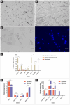
Figure 1
Figure 1
Chicken muscle cells. (A) Representative images of established chicken myoblasts derived from muscle tissues; scale bar 50 μm. (B) Representative images of established chicken myotubes obtained by myoblast differentiation; scale bar 50 μm. (C) Long tubular and multinucleated chicken myotube in bright field and counterstained with Hoechst 33342 (blue); scale bar 50 μm. (D) Relative gene expression of pluripotency genes (cSALL4, cSOX3, cKIT, cCLDN3, cOCT4, cLIN28A, and cNANOG), (E) muscle genes (cPAX7, cMYOD, cMYMK, and cMYH1E), and (F) extracellular matrix genes (cCollagen I α1, cCollagen I α2, cFibronectin, cLaminin, and cElastin) in primary cells isolated from chicken embryos. Data in (D–F) are shown as mean plus standard deviation. One asterisk indicates p ≤ 0.05, two asterisks indicate p ≤ 0.01 and three asterisks indicate p ≤ 0.001.
Figure 2
Figure 2
Immunofluorescence of chicken muscle cells. (A) Immunofluorescence staining of chicken myoblasts (B) and chicken myotubes with antibodies (green) paired box 7 (PAX7), myogenic factor 5 (MYF5), myogenic determination (MYOD), integrin alpha 7 (ITGA7), myogenin (MYOG), myosin heavy chain (MYHC), and desmin (DES). F-actin was counterstained with Rhodamine Phalloidin (orange), and nuclei were counterstained with Hoechst 33342 (blue). Scales bar 50 μm.
Figure 3
Figure 3
Adipogenic dedifferentiation of chicken mesenchymal stem cells (MSCs). Chicken MSCs were induced to dedifferentiate into adipocytes using L-α-Phosphatidylcholin and monitored over time. (A) At 0 h (day 0), undifferentiated MSCs displayed a fibroblast-like, spindle-shaped morphology typical of early-passage mesenchymal cells. (B) By day 4, the cells exhibited increased confluency and subtle morphological changes, with some adopting a more rounded shape indicative of early adipogenic commitment. At this early stage of induction, the cells are still proliferating and beginning to undergo morphological changes. The culture appears confluent as the MSCs maintain their fibroblast-like morphology and high proliferative capacity. (C) On day 6, a greater number of cells showed a rounded morphology along with the initial formation of intracellular vesicles, characteristic of early adipocytes. At this intermediate differentiation stage, some cells begin to round up and accumulate lipid droplets, a hallmark of early adipogenic commitment. During this transition, many cells detach or die, possibly due to their sensitivity to the induction medium or mechanical stress from media changes. This explains the apparent reduction in cell density compared to Panel B. Additionally, some lipid-filled cells may not adhere strongly to the surface, contributing to reduced confluency. (D) By day 11, cells had adopted a mature adipocyte-like phenotype, with prominent intracellular lipid droplets and a spherical shape. In the later stage of differentiation, the remaining adherent cells have adapted to the adipogenic conditions and completed differentiation. They exhibit robust lipid accumulation and a mature adipocyte-like phenotype. These cells tend to reoccupy the culture surface, and this contributes to the nearly confluent appearance seen here. Scales bar 50 μm.
Figure 4
Figure 4
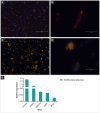
Dedifferentiated chicken adipocytes derived from mesenchymal stem cells. Fluorescent staining was used to visualize lipid accumulation and confirm adipogenic differentiation. (A) Dedifferentiated adipocytes stained with HCS LipidTOX™ Red Neutral Lipid Stain, showing widespread lipid droplet distribution throughout the culture. Nuclei were counterstained with Hoechst 33342 (blue). Scale bar: 300 μm. (B) Higher magnification of a single cell showing intracellular lipid droplets stained in red (LipidTOX) and nucleus in blue (Hoechst). Scale bar: 50 μm. (C,D) Cells stained with Nile Red, highlighting numerous intracellular lipid droplets as bright orange/yellow signals. Nuclei were counterstained with Hoechst 33342 (blue). Scale bars: 50 μm. (E) Relative gene expression of adipogenic markers (cPPARG, cFABP4, cADIPOQ, cADRP, and cPCK1) in dedifferentiated chicken adipocytes, confirming the adipogenic phenotype at the molecular level. Data are shown as mean plus standard deviation.
Figure 5
Figure 5
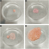
Chicken biomass constructs produced from muscle and adipose cells. (A) Muscle biomass produced from differentiated chicken myoblast appears compact, with a dense and uniform structure and a pink coloration typical of muscle tissue. (B) Adipogenic biomass generated from dedifferentiated chicken adipocytes presents a looser, more irregular morphology with a paler, translucent appearance, consistent with lipid-rich tissue. (C) Chicken muscle and adipose biomasses incorporating microcarriers are shown side by side, highlighting their morphological differences in structure, size, and consistency. (D) Combined muscle and adipose chicken biomass integrated with microcarriers, resulting in a larger, heterogeneous construct with a granular texture. The structure displays a mixture of pink and orange tones, reflecting the presence of both muscle and fat components, as well as the incorporated microcarriers that contribute to the bulk and support of the tissue.
Figure 6
Figure 6
Myoblast proliferation on commercial microcarriers over time in monolayer and spheroid cultures. (A) Cellva Ingredients (Brazil) microcarriers incubated without cells, showing the baseline structure and surface morphology. (B–D) Myoblasts cultured in monolayer on microcarriers. (B) After 24 hours of incubation, initial attachment of chicken myoblasts is observed on the microcarrier surface. (C) At 24 hours, a noticeable increase in cell coverage occurs, with myoblasts beginning to spread and form early connections. (D) By 96 hours, microcarriers are densely colonized by proliferating myoblasts, exhibiting extensive cell spreading and aggregation, indicating robust attachment and expansion. (E–G) Myoblasts cultured as spheroids on microcarriers. (E) At 1 hour, initial attachment of spheroid-associated cells begins. (F) By 24 hours, partial spreading and integration of spheroids with the microcarrier surface is visible. (G) At 96 hours, microcarriers exhibit large spheroid clusters, demonstrating strong aggregation and proliferation. Scale bar = 50 μm.

References

    1. Food, Nations (FAO) AO of the U (2023). Organization (WHO) WH Food safety aspects of cell-based food. Food safety aspects of cell-based food FAO; WHO.
    1. Feddern V, Bastos APA, Gressler V, Marques DMC, Ferreira FC, Rodrigues CAV, et al. Cell lines for cultivated meat production. Cultiv Meat. (2024):29–54. doi: 10.1007/978-3-031-55968-6_3 - DOI
    1. Bhat ZF, Bhat H, Pathak V. Prospects for in vitro cultured meat - a future harvest. Princ Tissue Eng Fourth Ed. (2013) 2013:1663–83. doi: 10.1016/B978-0-12-398358-9.00079-3 - DOI
    1. Ismail I, Hwang YH, Joo ST. Meat analog as future food: a review. J Anim Sci Technol. (2020) 62:111–20. doi: 10.5187/jast.2020.62.2.111, PMID: - DOI - PMC - PubMed
    1. Jairath G, Mal G, Gopinath D, Singh B. An holistic approach to access the viability of cultured meat: a review. Trends Food Sci Technol. (2021) 110:700–10. doi: 10.1016/j.tifs.2021.02.024 - DOI

LinkOut - more resources