Review
. 2025 Jul 14;13(1):101771.
doi: 10.1016/j.gendis.2025.101771. eCollection 2026 Jan.

The biomechanical signature of tumor invasion

Affiliations
Review

The biomechanical signature of tumor invasion

Chenhe Liu et al. Genes Dis. .

Abstract

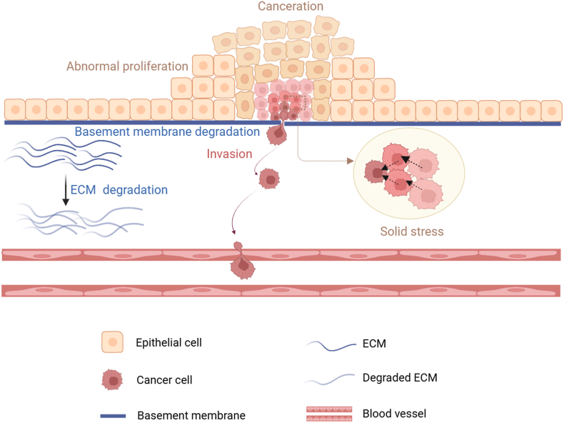
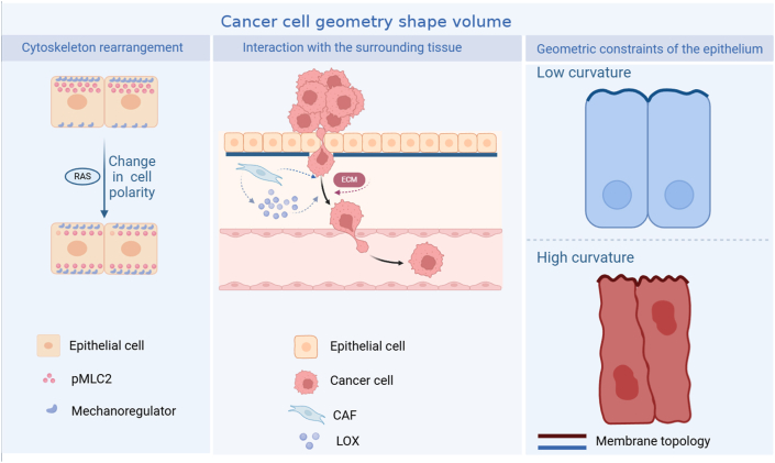
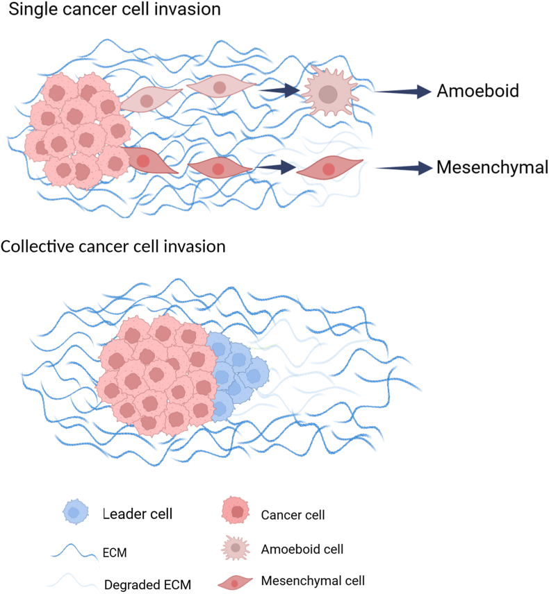
Tumor cell invasion is the key driver of metastatic dissemination, resulting in the development and progression of metastatic tumors at secondary sites, and remains the major cause of cancer-related death. Recent studies suggest that, in addition to protease-mediated degradation and chemotaxis-stimulated migration, tumor invasion is significantly influenced by physical surroundings. How tumor cells decode information about their shape deformation under mechanical stress and adapt their dynamic behavior to escape the confined regions remains largely unknown. This review highlights recent findings that illustrate mechanical cues in confined tumor microenvironment contribute to tumor progression. We also systematically discuss the role of compression-induced deformation in cell membrane topology and cytoskeletal remodeling, as well as its biophysical mechanisms in regulating tumor invasion from a biomechanical perspective.

Keywords: Actin remodeling; Mechanical forces; Mechanical memory; Microenvironment; Tumor invasion.


Conflict of interest statement

The authors declared no conflict of interests.

Figures

Figure 1
Figure 1
Tumor cell invasion process. As tumor cells abnormally proliferate and grow, they squeeze each other and undergo physical deformation because of mechanical forces such as solid stress. In the crowded microenvironment, tumor cells undergo epithelial-mesenchymal transition by secreting various cytokines and chemokines, at the same time, secreting metalloproteinases to degrade extracellular matrix (ECM) and promoting the invasion of tumor cells.
Figure 2
Figure 2
Architecture of tumor morphology. Proto-oncogenes-driven cytoskeletal rearrangements, geometric constraints of epithelial cells, and the tumor microenvironment jointly shape the morphological structure of tumor cells and their tissues., CAF, tumor-associated fibroblast; LOX, lysyl oxidase; pMLC2, phospho-myosin light chain 2.
Figure 3
Figure 3
Modes and mechanisms of tumor cell invasion. For single cell invasion, the characteristics of amoebic invasion are circular and highly deformed cell morphology, with bleb-like protrusions and Rho-associated kinase-dependent contraction of myosin. The lack of ability in intracellular adhesion and extracellular matrix (ECM) degradation weakens the interaction between cells and the surrounding matrix. The characteristics of mesenchymal invasion are slender cell morphology, with cytoskeletal contractility, integrin-mediated matrix adhesion, and peripheral protein hydrolysis. For collective cell invasion, it maintains high intracellular adhesion and front-rear polarity, relying on actin dynamics, integrin-mediated cell adhesion to ECM components, and ECM reorganization mediated by extracellular protein hydrolysis.
Figure 4
Figure 4
Biophysical traits of the tumor. The four major mechanical properties of tumors are solid stress (compression and tension), interstitial fluid pressure, matrix stiffness, and organizational microstructure. The physical and biological characteristics of the tumor interact synergistically.
Figure 5
Figure 5
The occurrence and development of benign and malignant breast cancer. When the benign breast cancer cells proliferate to a certain number, tumor cells undergo apoptosis, and the tumor eventually goes into decline after the different components of the immune system become active. For malignant breast cells with abnormal proliferation and infinite expansion, tumor cells adapt their dynamic behavior to escape the crowded physical surroundings by invasion and metastasis.,,
Figure 6
Figure 6
Extrusion direction determines the fate of tumor cells. Epithelia remove live or dead cells in response to cell stimulation caused by crowding or apoptosis, respectively. For Drosophila, a small part of anti-anoikis cells can overgrow within their lumen by apical extrusion, despite that most of them eventually die because of anoikis. Meanwhile, basal extrusion can lead to cell apoptosis. For vertebrates, basal extrusion can cause invasion of extruded cells, compared with apical extrusion-induced cell apoptosis.,
Figure 7
Figure 7
Surface topological morphology of human cervical epithelial cell carcinoma at different stages. Representative examples of 10 × 10 μm2 adhesion maps and height images of normal, pre-malignant, and malignant cells by atomic force microscopy. Normal cells look smoother, and malignant cells have more wrinkled surfaces in the adhesion. The brighter means the higher value of either adhesion or height.
Figure 8
Figure 8
The effect of cell membrane topology on tumor invasion. Epithelial cell carcinogenesis and abnormal proliferation induce cell crowding by physical compression, causing tumor cell deformation and changes in surface topology. The increased membrane curvature and the decreased membrane tension between the apical and basal sides drive cancerous epithelial cells to transform into invading amoebic or mesenchymal cells.
Figure 9
Figure 9
The proposed model describing how cell membrane topology and cortical actin act as the mechanical sensor of tumor invasion. As membrane curvature increases and tension decreases, the membrane topology changes from smooth to corrugated. At the same time, the ERM proteins connecting the plasma membrane and cortical actin dissociate from the membrane, and the BAR protein polymerizes from monomer/dimer to oligomers. Eventually, cortical actin filaments are converted into branched actin by the Arp2/3-WASP complex. Actin remodeling leads to tumor cell invasion by decreasing ezrin/radixin/moesin (ERM) proteins and assembling Bin/Amphiphysin/Rvs (BAR) proteins.
Figure 10
Figure 10
Molecular mechanisms of integrin-mediated actin remodeling in tumor invasion. The components of the actin complex in focal adhesions and invadopodia are similar, but their assembly, structure, and functions are completely different. Actin stress fibers are anchored to the plasma membrane at the integrin-dependent focal adhesions parallel to the extracellular matrix (ECM) (A), in contrast, the actin core in invadopodia is oriented perpendicular to the ECM and cell membrane (B). Invadopodia are built on a Tks5/Nck/WIP/N-WASP-dependent branched actin network, and Rho GTPase and Cdc42 drive pseudopodia-membrane protrusion, which is mediated by bundled actin filaments that form the actin core as the initial step in invadopodia assembly.

References

    1. Bray F., Laversanne M., Sung H., et al. Global cancer statistics 2022: GLOBOCAN estimates of incidence and mortality worldwide for 36 cancers in 185 countries. CA Cancer J Clin. 2024;74(3):229–263. - PubMed
    1. Githaka J.M., Pirayeshfard L., Goping I.S. Cancer invasion and metastasis: insights from murine pubertal mammary gland morphogenesis. Biochim Biophys Acta Gen Subj. 2023;1867(8) - PubMed
    1. Brabletz T., Kalluri R., Nieto M.A., Weinberg R.A. EMT in cancer. Nat Rev Cancer. 2018;18(2):128–134. - PubMed
    1. de Visser K.E., Joyce J.A. The evolving tumor microenvironment: from cancer initiation to metastatic outgrowth. Cancer Cell. 2023;41(3):374–403. - PubMed
    1. Raudenská M., Petrláková K., Juriňáková T., et al. Engine shutdown: migrastatic strategies and prevention of metastases. Trends Cancer. 2023;9(4):293–308. - PubMed

LinkOut - more resources