. 2025 Oct 31;11(44):eadw7879.
doi: 10.1126/sciadv.adw7879. Epub 2025 Oct 29.

The rise of lichens during the colonization of terrestrial environments

Affiliations

The rise of lichens during the colonization of terrestrial environments

Bruno Becker-Kerber et al. Sci Adv. .

Abstract

The origin of terrestrial life and ecosystems fundamentally changed the biosphere. Lichens, symbiotic fungi-algae partnerships, are crucial to nutrient cycling and carbon fixation today, yet their evolutionary history during the evolution of terrestrial ecosystems remains unclear due to a scarce fossil record. We demonstrate that the enigmatic Devonian fossil Spongiophyton from Brazil captures one of the earliest and most widespread records of lichens. The presence of internal hyphae networks, algal cells, possible reproductive structures, calcium oxalate pseudomorphs, abundant nitrogenous compounds, and fossil lipid composition confirms that it was among the first widespread representatives of lichenized fungi in Earth's history. Spongiophyton abundance and wide paleogeographic distribution in Devonian successions reveal an ecologically prominent presence of lichens during the late stages of terrestrial colonization, just before the evolution of complex forest ecosystems.


Conflict of interest statement

The authors declare that they have no competing interests.

Figures
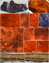
Fig. 1.
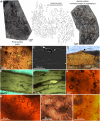
Fig. 1.. Morphology and internal structures of S. nanum.
(A) Isolated fragment of the thalli showing upper surface with pores. (B) Fragment under transmitted light. (C and D) Anastomosing hyphae. (E) Hypha with septum (sep). (F) Stereomicroscope image of the upper surface with open (op) and closed pores (cp). (G) Hyphae associated with and branching toward photobiont cells and cell packages. (H) Putative asci- or conidium-like structures (white arrows) and possible spores (black arrow). (I) Cells and cell packages. (J and K) Swollen cells (sc). (L and M) Thin sections with longitudinal cuts illustrating the outermost upper layer (ol) on the upper surface.
Fig. 2.
Fig. 2.. Thalli disposition and internal cellular structures.
(A) Slab with S. nanum connected thalli elements showing their distribution on the bedding plane. (B) Optical microscope image showing open and closed pores (arrow). (C) Slice from SR-nanoCT in the region of an open pore showing remnants of the former cover. (D) Poral covering remnants observed by transmitted light. (E and F) Bright yellow algae and dark yellowish green thalli under reflected fluorescence light. (G to J) Photobiont cells and cell packages strongly associated with hyphae networks.
Fig. 3.
Fig. 3.. SR-nanoCT 3D surface rendering of S. nanum.
(A) External surface showing an open pore. (B and C) Translucent image of the same sample with the distribution of internal structures. (D) Internal photobiont cells (green) and hyphae (white). (E) Plane view of internal hyphae in another specimen with photobiont cells (green). (F) Perpendicular view of (E). Scale bars, 500 μm (A), 400 μm [(B) to (D)], 100 μm (E), and 50 μm (F).
Fig. 4.
Fig. 4.. SR-nanoXRF and microFTIR chemical maps showing calcite particles and calcite-preserved hyphae and Zn microparticles.
(A to E) SR-nanoXRF of a longitudinal section of S. nanum, perpendicular to its surface, showing Ca (A), Fe (B), and Zn (C) maps, and respective insets for Ca (D) and Zn (E). (F) MicroFTIR map in the carbonate stretching vibration region. us, upper surface; hy, hyphae.
Fig. 5.
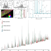
Fig. 5.. Chemical composition of S. nanum.
(A) deconvolution of transmission FTIR spectra in the region of C═O, C═C, and C─N. (B) Fitting of N1s XPS spectrum. (C) Superposed MicroFTIR chemical maps. (D) Spectra from the points arrowed in (C), showing differences in the C─N/N─H (green) and C═O (red) integrated areas. au, arbitrary units. (E) ToF-SIMS spectra in negative ion mode with peaks assigned to N-containing fragments. (F) GC-MS full-scan chromatogram of the polar fraction of S. nanum pyrolysate highlighting the relative abundances of straight-chain alkyl pyridines, alcohols, and ketones.
Fig. 6.
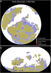
Fig. 6.. Paleogeographic distribution of Spongiophyton by the Early Devonian.
(A) 3D orthographic plot with color-coded occurrences and the plate reconstruction model from ref (84). Colors from red to pale orange represent older to younger relative ages. Grey-colored circles represent less well-defined ages. (B) Same distribution as in (A) but in a Robinson projection.
Fig. 7.
Fig. 7.. Artistic reconstruction of Spongiophyton during the Early Devonian in the high latitude depositional system of the Paraná Basin.
Paleoart by J. Lacerda.

References

    1. Strother P. K., Servais T., Vecoli M., The effects of terrestrialization on marine ecosystems: The fall of CO2. Geol. Soc. Lond. Spec. Publ. 339, 37–48 (2010).
    1. Edwards D., Cherns L., Raven J. A., Could land-based early photosynthesizing ecosystems have bioengineered the planet in mid-Palaeozoic times? Palaeontology 58, 803–837 (2015).
    1. Rubinstein C. V., Gerrienne P., de la Puente G. S., Astini R. A., Steemans P., Early Middle Ordovician evidence for land plants in Argentina (eastern Gondwana). New Phytol. 188, 365–369 (2010). - PubMed
    1. Spaak G., Edwards D. S., Foster C. B., Pagès A., Summons R. E., Sherwood N., Grice K., Environmental conditions and microbial community structure during the Great Ordovician Biodiversification Event; a multi-disciplinary study from the Canning Basin, Western Australia. Glob. Planet. Change 159, 93–112 (2017).
    1. Libertín M., Kvaček J., Bek J., Žárský V., Štorch P., Sporophytes of polysporangiate land plants from the early Silurian period may have been photosynthetically autonomous. Nat Plants 4, 269–271 (2018). - PubMed

LinkOut - more resources