. 2025 Oct 29;15(1):37866.
doi: 10.1038/s41598-025-21680-5.

A high-throughput drug screening assay for anti-tau aggregation using split GFP and flow cytometry

Affiliations

A high-throughput drug screening assay for anti-tau aggregation using split GFP and flow cytometry

Omnia M H Ibrahium et al. Sci Rep. .

Abstract

Tau protein aggregation is a hallmark of neurodegenerative diseases, including Alzheimer's disease, making the development of anti-aggregation therapeutics a critical area of research. Progress in drug discovery has been hindered by the lack of efficient screening methods that accurately reflect cellular conditions. We present a high-throughput cell-based assay utilizing split GFP technology to monitor tau aggregation in living cells. Our system employs suspension-adapted HEK293 cells co-transfected with tau proteins fused to complementary GFP fragments, producing fluorescent signals upon tau aggregation. Notably, our system demonstrates tau aggregation without external aggregation inducers, likely due to the enhanced protein expression in suspension-adapted cells. Validation with a known urea-based tau aggregation inhibitor showed dose-dependent reduction in fluorescence, corresponding to decreased tau aggregation. The assay's flow cytometry compatibility enables rapid, quantitative analysis of large sample sets while allowing simultaneous assessment of compound efficacy and cytotoxicity. This method advances tau aggregation monitoring and drug discovery by providing a physiologically relevant platform for identifying novel anti-tau aggregation therapeutics.


Conflict of interest statement

Declarations. Competing interests: The authors declare no competing interests.

Figures

Fig. 1
Fig. 1
The assay employs split GFP fragments (GFP10 and GFP11) fused to 0N4R Tau to detect Tau aggregation in living cells. Co-transfection of Tau-GFP10 and Tau-GFP11 constructs leads to protein expression. Upon Tau aggregation, the GFP fragments reconstitute a functional GFP, producing a fluorescent signal. The assay can be scaled to microtiter plates for transfection, cell growth, and the addition of potential Tau aggregation inhibitors. The fluorescent signal is quantified using high-throughput methods such as flow cytometry, enabling efficient screening of candidate compounds that prevent or reduce Tau aggregation.
Fig. 2
Fig. 2
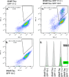
A and B: HEK293 cells transfected individually with GFP10-Tau (A) or GFP11-Tau (B) show negligible GFP fluorescence, confirming that neither split GFP fragment fluoresces on its own. (C) Co-transfection of cells with GFP10-Tau and GFP11-Tau results in a significant increase in GFP fluorescence, indicative of Tau–Tau interactions leading to GFP reconstitution. (D) Concatenated histogram comparing fluorescence profiles of individual transfectants (gray, Total+) and co-transfected cells (green, GFP+), highlighting the GFP-positive population resulting from Tau aggregation.
Fig. 3
Fig. 3
Time course of GFP expression in transfected cells using the split GFP Tau aggregation assay.
Fig. 4
Fig. 4
Analysis of drug candidate effect on tau aggregation and cell viability. (A) Concatenated histogram and scatter plot showing GFP fluorescence gated on live cells (GFP+, green) and total cell population (Total+, blue) across increasing concentrations of drug treatment in Expi293 cells co-transfected with GFP10-Tau and GFP11-Tau constructs. The 0 μM condition represents the DMSO vehicle control (0.1% final concentration), used in all treatment groups. (B) Nonlinear regression analysis of the dose-response curve using a four-parameter logistic model. The Hill slope was − 0.6554, indicating mild negative cooperativity in the inhibitory response. (C) Initial reduction in cell viability was observed with DMSO vehicle control compared to untreated cells. (D) Drug treatment showed significant cytotoxicity (p < 0.0001) only at concentrations above 30 μM when compared to DMSO-treated cells. (E) Western blot analysis revealed a reduction in the intensity of the ~108 kDa band corresponding to the Tau-GFP10C/Tau-GFP11C complex in urea based compund-treated samples, supporting the observed inhibition of Tau aggregation. with either DMSO, urea compound or no treatment. GAPDH was used as a loading control. Full-length, uncropped blots corresponding to this figure are provided in Tau WB Images.pptx Supplementary Files. (F) Comparison of drug addition time points post-transfection. Drug compounds added at 3 h post-transfection demonstrated a more pronounced inhibition of Tau aggregation compared to those added at 24 h post-transfection.
Fig. 5
Fig. 5
Fluorescent microscopy images of HEK293T adherent cells and suspension-adapted Expi(HEK)293 cells co-transfected with GFP10-Tau and GFP11-Tau constructs. Scale bars indicate 100 μm.

References

    1. Iqbal, K., Liu, F., Gong, C-X., Alonso, A. D. C. & Grundke-Iqbal, I. Mechanisms of tau-induced neurodegeneration. Acta Neuropathol. (Berl.)118, 53–69. 10.1007/s00401-009-0486-3 (2009). - PMC - PubMed
    1. Lasagna-Reeves, C. A. et al. Tau oligomers impair memory and induce synaptic and mitochondrial dysfunction in wild-type mice. Mol. Neurodegener.6, 39. 10.1186/1750-1326-6-39 (2011). - PMC - PubMed
    1. Buée, L., Bussière, T., Buée-Scherrer, V., Delacourte, A. & Hof, P. R. Tau protein isoforms, phosphorylation and role in neurodegenerative disorders. Brain Res. Rev.33, 95–130. 10.1016/S0165-0173(00)00019-9 (2000). - PubMed
    1. Biernat, J., Gustke, N., Drewes, G., Mandelkow, E. & Mandelkow, E. Phosphorylation of Ser262 strongly reduces binding of Tau to microtubules: Distinction between PHF-like immunoreactivity and microtubule binding. Neuron11, 153–163. 10.1016/0896-6273(93)90279-Z (1993). - PubMed
    1. Trojanowski, J. Q. & Lee, V. M. Phosphorylation of paired helical filament Tau in alzheimer’s disease neurofibrillary lesions: Focusing on phosphatases. FASEB J.9, 1570–1576. 10.1096/fasebj.9.15.8529836 (1995). - PubMed

MeSH terms

LinkOut - more resources