. 2025 Oct;646(8087):1096-1104.
doi: 10.1038/s41586-025-09650-3. Epub 2025 Oct 29.

Ultrasound-driven programmable artificial muscles

Affiliations

Ultrasound-driven programmable artificial muscles

Zhan Shi et al. Nature. 2025 Oct.

Abstract

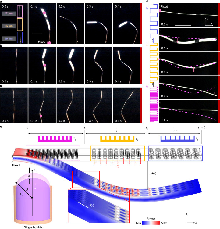
Muscular systems1, the fundamental components of mobility in animals, have sparked innovations across technological and medical fields2,3. Yet artificial muscles suffer from dynamic programmability, scalability and responsiveness owing to complex actuation mechanisms and demanding material requirements. Here we introduce a design paradigm for artificial muscles, utilizing more than 10,000 microbubbles with targeted ultrasound activation. These microbubbles are engineered with precise dimensions that correspond to distinct resonance frequencies. When stimulated by a sweeping-frequency ultrasound, microbubble arrays in the artificial muscle undergo selective oscillations and generate distributed point thrusts, enabling the muscle to achieve programmable deformation with remarkable attributes: a high compactness of approximately 3,000 microbubbles per mm2, a low weight of 0.047 mg mm-2, a substantial force intensity of approximately 7.6 μN mm-2 and fast response (sub-100 ms during gripping). Moreover, they offer good scalability (from micrometre to centimetre scale), exceptional compliance and many degrees of freedom. We support our approach with a theoretical model and demonstrate applications spanning flexible organism manipulation, conformable robotic skins for adding mobility to static objects and conformally attaching to ex vivo porcine organs, and biomimetic stingraybots for propulsion within ex vivo biological environments. The customizable artificial muscles could offer both immediate and long-term impact on soft robotics, wearable technologies, haptics and biomedical instrumentation.


Conflict of interest statement

Competing interests: The authors declare no competing interests.

Figures

Fig. 1
Fig. 1. Ultrasound-actuated microbubble-array artificial muscles.
a, A uniform-size microbubble-array artificial muscle consists of thousands of microbubbles on its bottom surface. Under continuous ultrasound excitation, the artificial muscle bends upwards with different excitation voltages, labelled as V1, V2 and V3. Inset: the input ultrasound signal with modulated amplitude versus time. b, A variable-size microbubble-array artificial muscle comprises three microbubble arrays with different diameters (d), each corresponding to a distinct natural frequency (f) and represented by the colours purple, yellow and grey. c, Under sweeping-frequency ultrasound excitation, the artificial muscle exhibits multimodal deformation in the time domain, shown at time points T1, T2 and T3. d, Schematic of a soft gripper constructed with an array of artificial muscles patterned with uniform-size microbubble arrays. Upon ultrasound excitation, these muscles close simultaneously in milliseconds. e, Schematic of a bioinspired stingraybot incorporating variable-size microbubble-array artificial muscles. Under sweeping-frequency ultrasound excitation, the stingraybot enacts undulating propulsion. f, A silicon wafer with micropillar arrays serves as the negative mould of microbubble cavities in standard soft-lithography fabrication. Inset: the micropillar array. g, A prototype of the stingraybot near a 5-cent Swiss franc coin. h, Left: trapped microbubble arrays. Right: upwards microstreaming jets generated from a microbubble array oscillating under ultrasound excitation visualized by 6-μm-diameter tracer microparticles. n = 3 independent samples. Scale bars, 300 μm (f, inset), 2 cm (g), 500 μm (h, left), 100 μm (h, right).
Fig. 2
Fig. 2. Actuation and modelling of microbubble-array artificial muscles.
ac, Time-lapse images of the selective deformation shapes of a variable-size microbubble-array artificial muscle (3 cm × 0.5 cm × 80 μm) containing microbubbles of diameter 12 μm, 16 μm and 66 μm, each 50 μm in depth, excited at 96.5 kHz (a), 82.3 kHz (b) and 33.2 kHz (c), respectively, at 60 VPP. The pink dots indicate the region of the bubble array being activated. d, Time-lapse images of the variable-size microbubble-array artificial muscle under sweeping-frequency ultrasound excitation (20–90 kHz, 1.2 s, 60 VPP). The pink dashed lines mark the shape of the muscle at the previous time step and the pink arrows mark the bending direction of the excited part. e, Modelling of the activation mechanism of microbubble-array artificial muscles. The pink, yellow and blue boxes represent differently sized microbubble-array segments. The upper portion illustrates schematics of the cross-section of the artificial muscle, each part of the artificial muscle corresponding to a specific length (L) and second moment of area (I). Fi denotes the thrust force generated by the microstreaming (here the yellow segment of the muscle generates thrust), Δ and θ denote the deflection and rotation angle along the long axis (x axis), and s denotes the coordinate along the beam. Lower-left inset: modelling of a microbubble, where Rc is the radius of the cavity, R is the curvature radius of the trapped microbubble and a is the amplitude of the centre displacement during oscillation. Scale bars, 1 cm (a,d).
Fig. 3
Fig. 3. Adaptive gripper and robotic skin based on microbubble-array artificial muscles.
a, Time-lapse sequence showing a live zebrafish larva grasped by a soft gripper composed of multiple artificial-muscle petals (10 mm × 0.7 mm × 80 µm), each incorporating microbubble arrays (12 µm in diameter × 50 µm in depth). Inset: magnified view of the larva. b, Rotation of an almond by a conformable microbubble-array robotic skin (12 μm × 50 μm). c, Deformation of a blade of grass by the same robotic skin, showing self-attachment and actuation. Inset: magnified view of the microbubble array. d, Conformal attachment of a green fluorescently labelled cardiac patch (30 mm × 10 mm × 80 μm) to the epicardial surface of an ex vivo porcine heart. e, Experimental set-up showing an excised porcine bladder with an ultrasound transducer positioned approximately 5 cm from the left side and an endoscope inserted for internal visualization. f, Time-lapse endoscopic images showing the encapsulated artificial muscle inside the bladder, its release at approximately 3–5 min and conformal attachment to the inner wall at 4.2 min under ultrasound activation. Scale bars, 5 mm (ac), 1 cm (d,f), 2 cm (e).
Fig. 4
Fig. 4. Bioinspired swimming and navigation within ex vivo biomedical environment.
a, Undulatory motion of the microbubble-array fins (12 μm, 16 μm and 66 μm in diameter, 50 μm in depth) of the bioinspired stingraybot before release. b, Forward swimming of the stringraybot under sweeping-frequency excitation (30–90 kHz, 2 s, 60 VPP). Right: fin motion during swimming. Lower inset: schematic of the patterned microbubble arrays. In a and b, the pink dashed lines and arrows denote the fin shapes in last step and the fin’s moving direction, respectively. c, Edible hydroxypropyl methylcellulose capsule (27 mm × 12 mm) containing a pre-folded stingraybot. d, Set-up for release and navigation of the encapsulated artificial muscle in an excised porcine stomach, with an external transducer positioned approximately 3 cm from the stomach and internal endoscope for visualization. e, Locomotion of the stingraybot inside an excised porcine stomach. f, Locomotion of a pre-folded, wheel-shaped artificial muscle (30 mm × 5 mm × 80 μm) with variable-size microbubble arrays (12 μm, 16 μm and 66 μm in diameter, 50 μm in depth) inside a porcine stomach. The artificial muscle propels along the stomach surface under sweeping-frequency excitation (30–100 kHz, 2-s sweep period, 60 VPP). The blue arrows mark the direction of motion and the green dots indicate the centre position. Inset: pre-folded shape. g, Set-up for ex vivo manipulation of a pre-folded artificial muscle inside an excised porcine intestine, with external transducers and an internal endoscope. Inset: endoscopic view of the artificial muscle. h, Time-lapse images showing the artificial muscle rolling along the curved mucosal wall under ultrasound sweeping-frequency (30–100 kHz, 2-s sweep period, 60 VPP) delivered by a piezo transducer. i, Locomotion of the artificial muscle driven by a high-intensity focused ultrasound transducer (1–3 MHz, 1-s sweep period, 60 VPP). Red lines, trajectory; yellow dots, centre position over time. Scale bars, 1 cm (ac,e,f,h,i), 2 cm (d,g).
Extended Data Fig. 1
Extended Data Fig. 1. Characterization of the resonance frequencies of differently sized microbubbles.
a, Oscillation amplitude of a 60 μm × 150 μm microbubble as a function of the ultrasound excitation frequency. According to the measured oscillation amplitude of the microbubbles, the resonance frequency is identified to be 35.5 kHz. b, Experimentally measured resonance frequencies of microbubbles with depths of 150 μm and 175 μm, radii ranging from 20 to 70 μm in 5 μm increments, and a depth of 50 μm with radii of 6 μm, 8 μm, 20 μm, 30 μm, 33 μm and 40 μm. The solid lines represent numerically predicted results. Additionally, notable discrepancies exist between the calculated and measured resonance frequencies, which may originate from transducer coupling with the glass slide, interactions between bubbles, or other factors.
Extended Data Fig. 2
Extended Data Fig. 2. Selective actuation of microbubble arrays.
a, Microstreaming respectively generated by 40 μm, 60 μm, and 80 μm microbubble arrays under ultrasound frequencies of 76.3 kHz (green), 57.4 kHz (blue), and 27.6 kHz (purple). Colored boxes indicate the activated microbubbles. The black dashed line denotes the measurement position of the microstreaming velocity, which is 100 μm away from the surface. b, Measured streaming flow of a with 2 μm tracer microparticles. The streaming flow field was analyzed by PIV (Matlab R2022b, PIVlab 2.60). The color bar denotes the particle moving velocity perpendicular to and extending away from the bubble surface. c, Plot of the measured microstreaming velocity along the long axis of the variable-size microbubble array under different excitation frequencies. d, Simulations of microstreaming around the bubble array with three different excitation frequencies. The pink dotted lines in a, b, and d delineate the boundaries of three distinct microbubble arrays.
Extended Data Fig. 3
Extended Data Fig. 3. Measurement of microstreaming velocity by PIV.
a, Microstreaming generated by a 4×4 80 μm × 150 μm microbubble array under an excitation frequency of 27.6 kHz and two different voltages of 10 VPP (left panel) and 30 VPP (right panel). The black dashed line denotes the measurement position of the microstreaming velocity, which is 80 μm away from the surface. b, Plot of microstreaming velocity versus ultrasound excitation voltage respectively measured by 4×4 microbubble arrays with three different sizes (40 μm, 60 μm, and 80 μm). The solid lines are the quadratic fitting results. The shaded error bands represent mean ± s.d. from n = 5 independent measurements.
Extended Data Fig. 4
Extended Data Fig. 4. Attachment of a robotic patch to an ex vivo porcine heart.
a, Experimental setup showing an artificial muscle positioned between an ultrasound transducer (operated at 96 kHz and 60 VPP) and the heart, separated by ~2.5 cm. b, The muscle is released from the tweezer positioned at the bottom and rises upward due to buoyancy (trajectory indicated by the red arrow). c, The artificial muscle conforms to the surface of the heart when stimulated by ultrasound (trajectory indicated by the red arrow). d, Time course of attachment robustness, demonstrating conformation and adhesion of the artificial muscle to the heart from 0 to 60 min under ultrasound actuation, followed by detachment upon ultrasound deactivation at 70 min. The pink dashed rectangle indicates the location of the artificial muscle.
Extended Data Fig. 5
Extended Data Fig. 5. Multimodal shape transformation of a microbubble array-patterned functional surface.
a, Multimodal shape transformation of a circular surface under continuous ultrasound excitation frequencies of 96.2 kHz, 82.5 kHz, and 33.2 kHz, respectively. The circular surface was topped with a circular PDMS block (2 cm diameter, 0.3 cm thickness) to reduce buoyancy. The inset shows a schematic of the microbubble array patterned on the surface. b, Dynamic shape transformation of the circular surface under sweeping-frequency ultrasound excitation spanning from 10 kHz to 100 kHz over 2 s.
Extended Data Fig. 6
Extended Data Fig. 6. Ultrasound-enhanced dye delivery into an agar phantom by a microbubble-array robotic patch.
a, Setup illustrates an agar block resting in an acoustic tank, with a piezo transducer positioned 5 cm to the left. A circular robotic patch is placed on the agar block with its microbubble arrays facing downward. The zoom–in highlights the patch surface; inset shows the patterned microbubble array. b, Agar block prior to dye exposure. c, Control condition showing the agar block after 30 min in a dye-filled tank without ultrasound actuation. d, Top and cross-sectional views of the agar block after 30 min of ultrasound actuation (96 kHz, 60 VPP), revealing enhanced dye penetration. The blue dashed line indicates the cutting plane; green dashed lines delineate the boundaries of the penetrated region.
Extended Data Fig. 7
Extended Data Fig. 7. Artificial muscles functioning at scales from 10−1 mm to 102 mm.
a, A microscale rotator featuring an asymmetric 8 × 8 microbubble array (12 μm × 50 μm). The upper and lower panels show the microrotator with ultrasound off and on, respectively, at 95.5 kHz and 60 VPP. n = 3 independent measurements. b, A millimeter-scale artificial muscle with an asymmetric 400 × 200 microbubble array (12 μm × 50 μm). The upper and lower panels show the device with ultrasound off and on, respectively, under the same driving conditions. The purple dashed line indicates the original position of the artificial muscle. c, A macroscale stingraybot equipped with artificial muscles comprising variable-size microbubble arrays (40 μm × 150 μm, 60 μm × 150 μm, 80 μm × 150 μm, respectively), demonstrating undulatory motion under excitation (10–90 kHz, duty cycles 2 s, 120 VPP). The blue line marks the current location of the fin edge, while the white dashed line shows its position in the previous frame of the time-lapse image.
Extended Data Fig. 8
Extended Data Fig. 8. Deformation of uniform-size microbubble array artificial muscle.
a, Time-lapse images of a uniform-size microbubble array artificial muscle with microbubbles respectively positioned at the left and right side. The pink rectangle and arrow show the fixed end and bending direction of the muscle, respectively. The red line on the left side denotes the location of the transducer. b, Plot of the bending amplitude of the muscle tip over multiple excitation cycles with an average repeated-positioning error of ±0.8 mm with the excitation signal shown in the top panel. The pink and gray dots correspond to the excitation ‘on’ (80.5 kHz and 52.5 VPP) and ‘off’ stages, respectively. The green and blue dots represent the measured deformation amplitudes when the bubbles are positioned on the left and right sides, respectively, as shown in a. c, Bending amplitude of uniform-size microbubble array artificial muscles with microbubble diameters (D) of 40 μm, 60 μm, and 80 μm and without microbubbles under excitation voltages from 1.5 to 52.5 VPP. The microbubbles have a constant depth of 50 μm. The dots and solid lines are the experimental results and quadratic fitting results, respectively. The shaded error bands represent mean ± s.d. from n = 5 independent measurements. All the muscles bent in the direction as shown in the left panel of a.
Extended Data Fig. 9
Extended Data Fig. 9. Ultrasound-induced deformation of an artificial muscle in porcine blood.
An artificial muscle embedded with uniform microbubbles (12 μm × 50 μm) was immersed in 100% porcine blood, with a piezoelectric transducer bonded to the bottom of the acoustic tank and positioned 3 cm from the muscle. Final deformation (|Δ|) of the artificial muscle as a function of excitation amplitude—0.44 cm, 0.27 cm, 0.10 cm and 0.04 cm corresponding to 60 VPP, 45 VPP, 30 VPP and 15 VPP, respectively—under excitation at 96 kHz. Yellow dashed lines indicate the initial muscle position; white dashed lines indicate the deformed position.
Extended Data Fig. 10
Extended Data Fig. 10. Artificial muscles performance comparison.
a, Response time versus gripping ability (LO/LG) for grippers using different actuation methods, where LO and LG are the dimensions of the gripped object and gripper, respectively. b, Comparison of force-to-weight ratios of grippers versus their size for various actuation methods. c, Comparison of relative swimming speeds (body lengths per second) of swimmers with different actuation mechanisms across various scales (microscale to macroscale). Shaded regions (convex hulls) indicate typical performance ranges; representative studies are labeled by author and year.

References

    1. Gordon, M. S. et al. Animal Locomotion: Physical Principles and Adaptations (CRC Press, 2017).
    1. Li, M., Pal, A., Aghakhani, A., Pena-Francesch, A. & Sitti, M. Soft actuators for real-world applications. Nat. Rev. Mater.7, 235–249 (2022). - PMC - PubMed
    1. Zhang, J. et al. Robotic artificial muscles: current progress and future perspectives. IEEE Trans. Robot.35, 761–781 (2019).
    1. Laschi, C., Mazzolai, B. & Cianchetti, M. Soft robotics: technologies and systems pushing the boundaries of robot abilities. Sci. Robot.1, eaah3690 (2016). - PubMed
    1. Rus, D. & Tolley, M. T. Design, fabrication and control of soft robots. Nature521, 467–475 (2015). - PubMed

LinkOut - more resources