. 2025 Oct 28;13(11):e71139.
doi: 10.1002/fsn3.71139. eCollection 2025 Nov.

Combining Network Pharmacology, Molecular Docking, and Integrative Studies to Explore the Mechanism of Helminthostachys zeylanica in Alleviating Ulcerative Colitis

Affiliations

Combining Network Pharmacology, Molecular Docking, and Integrative Studies to Explore the Mechanism of Helminthostachys zeylanica in Alleviating Ulcerative Colitis

Chih-Ting Lin et al. Food Sci Nutr. .

Abstract

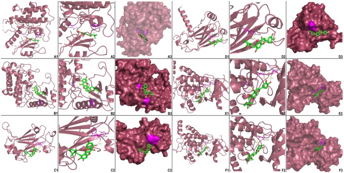
Helminthostachys zeylanica (L.) hook (HZ), has recently gained attention as a potential herbal supplement for managing ulcerative colitis (UC) through its bioactive compounds. To comprehensively investigate HZ's therapeutic effects and underlying mechanisms on UC, we utilized network pharmacology and in vitro and in vivo analyses. The therapeutic potential of HZ was evaluated using a DSS-induced mouse model of ulcerative colitis, alongside in vitro cellular studies. A network pharmacology approach was first used to predict the active compounds and molecular targets of HZ. Subsequently, integrated experimental techniques-including ELISA, Western blotting, histological analysis, immunofluorescence, flow cytometry, and molecular docking-were employed to validate and support the predicted mechanisms. Network pharmacology analysis identified 15 active compounds in HZ, contributing to its multi-target synergistic activity and anti-inflammatory effects. HZ was found to modulate multiple inflammatory pathways, particularly the Toll-like receptor 4 (TLR4) and NF-κB signaling pathways, regulating vital inflammatory mediators such as tumor necrosis factor-α (TNF-α), interleukin-6 (IL-6), and interleukin-1β (IL-1β), emphasizing its therapeutic potential in UC. ELISA, Western blot, and histological analyses confirmed that HZ significantly reduced colon inflammation. Immunofluorescence and flow cytometry analyses also demonstrated that HZ alleviated inflammation by regulating TLR4/NF-κB and CD3 signaling pathways without involving apoptosis. Ultimately, molecular docking further identified core compounds, including Ugonin M, O, K, and R, which exhibited strong binding affinity to critical proteins in the TLR4/NF-κB pathway, such as TAK1, IKKβ, and RELA, underscoring their role in HZ's anti-inflammatory mechanisms. Collectively, these findings provide a solid basis for further investigation into the mechanistic effects and broader clinical potential of HZ as a therapeutic approach for UC.

Keywords: Helminthostachys zeylanica; TLR4/NF‐κB signaling pathway; network pharmacology; ugonin; ulcerative colitis.


Conflict of interest statement

The authors declare no conflicts of interest.

Figures

FIGURE 1
FIGURE 1
(A) Components‐compounds‐targets network of HZ. The green hexagons represent the 18 active compounds derived from HZ, while the pink circles denote the 99 potential target genes. The nodes' sizes indicate the active compounds' DC, with other target gene nodes displayed in a standardized size. (B) The interactive PPI network of HZ target proteins associated with UC. The size of the nodes is positively correlated with the node degree criterion. The varying width and transparency of the edges, determined by the combined score of two target proteins, illustrate the extent of their interactions. Additionally, proteins with similar biological functionalities are grouped into distinct clusters.
FIGURE 2
FIGURE 2
(A) The GO enrichment annotation of target genes shows the functional distribution across BP, CC, and MF terms. These terms were ranked based on gene counts, p‐values, and fold enrichment. Different shapes indicate distinct GO terms. The y‐axis represents gene functional classifications, while the x‐axis shows the corresponding fold enrichment values. There is a positive correlation between node size and the number of target genes, and the color gradient corresponds to the negative logarithm of the p‐value, with brighter red shades indicating higher values. (B) KEGG enrichment analysis of target genes. The color scale reflects varying thresholds of adjusted p‐values, while the dot sizes represent the gene count for each pathway. The y‐axis categorizes the KEGG pathways, and the x‐axis displays the level of fold enrichment.
FIGURE 3
FIGURE 3
(A) Weight loss monitoring of mice. (B) Average DAI score of mice. (C) The colon length of each group on day 12 is presented. The data represent four experiments, with scale bars indicating 1 cm. Results are expressed as means ± standard error (n = 5). *p < 0.01 versus control mice; #p < 0.05 versus DSS‐treated mice. (D) Representative H&E and PAS staining of colon tissue (magnification, ×200) is shown. Abbreviations used in microscopic images include colonocytes, goblet cells, crypts, lamina propria, mucosa, submucosa (SM), and muscularis mucosa (MM). Key highlights include the disappearance of colonocytes (bold arrows) and the infiltration of inflammatory substances and neutrophils (stars).
FIGURE 4
FIGURE 4
(A) TNF‐α concentration in the plasma of mice from all groups. *p < 0.05 versus DSS mice; **p < 0.01 versus DSS mice. (B) IL‐6 concentration in the colon and intestinal tissues of mice from all groups. *p < 0.01 (colon tissue); #p < 0.05 (intestinal tissue). (C) TNF‐α concentration in the medium of IEC‐6 and T84 cells from all groups. (D) IL‐6 concentration in the medium of IEC‐6 and T84 cells from all groups. (E) Immunofluorescent staining with CD3 in colon tissues from mice. Red staining indicates CD3, while DAPI (blue) is a nuclear stain. All images are magnified ×400.
FIGURE 5
FIGURE 5
(A) Immunofluorescent staining with TLR4 in colon mucosa from mice. Immunofluorescent staining for TLR4 (red) staining was performed in the inflamed colon. Nuclei are stained with blue (DAPI); all magnifications are × 400. (B) Western blot analysis of TLR4 (95 kDa), NF‐κB p65 (RELA, 65 kDa), and Actin (42 kDa) in colon tissue. (C) The relative intensity of each band in the western blots was determined using densitometry, and the ratios were calculated. *p < 0.05 versus DSS mice in TLR4 expression.
FIGURE 6
FIGURE 6
(A) Flow cytometry analysis on cell cycle of T84 treated with HZ of different concentrations for 16 h. (B) Cell cycle distribution of T84 cells.
FIGURE 7
FIGURE 7
3D molecular docking visualization results of target proteins with active compounds of HZ (A) Ugonin M with MAP3K7 (5JGA). (B) Ugonin O with MAP3K7 (5JGA). (C) Ugonin M with IKBKB (4KIK). (D) Ugonin R with IKBKB (4KIK). (E) Ugonin K with RELA (1IKN). (F) Ugonin O with RELA (1IKN). Green‐red stick models represent active compounds, while the secondary structure of the proteins is depicted using dark burgundy ribbon representations. The yellow lines between active compounds and target proteins indicate the polar hydrogen bonding interactions. Additionally, surface conservation analysis highlights how receptor compounds bind to ligand proteins.
FIGURE 8
FIGURE 8
The mechanism of HZ in UC involves the TLR4/NF‐κB pathway. The anti‐inflammatory properties of HZ on UC are mediated through the TLR4/NF‐κB signaling pathway, encompassing the inhibition of cytokine production and prevention of T cell activation and differentiation. HZ achieves a downregulation of TLR4 and NF‐κB, resulting in reduced cellular release of TNF and IL‐6. Subsequent molecular docking simulation analysis reveals that the pure compounds of HZ, namely UgoninM, O, R, and K, exert their inhibitory effects on the TLR4/NF‐κB signaling pathway by influencing proteins such as TAK1, IKKβ, and RELA. Consequently, this impedes the degradation of the NF‐κB trimer, preventing its entry into the cell nucleus and activating pro‐inflammatory effects. On another note, CD3+ T cells, when associated, play a role in enhancing the production of specific antibodies by immune cells through the secretion of cytokines such as IL‐6 and TNF, thereby amplifying the extent of humoral immune responses.

References

    1. Abreu‐Martin, M. T. , and Targan S. R.. 1996. “Chapter 18—Lamina Propria Lymphocytes: A Unique Population of Mucosal Lymphocytes.” In Essentials of Mucosal Immunology, edited by Kagnoff M. F. and Kiyono H., 227–245. Academic Press. 10.1016/B978-012394330-9/50020-X. - DOI
    1. Andersen, V. , Christensen J., Ernst A., et al. 2011. “Polymorphisms in NF‐κB, PXR, LXR, PPARγ and Risk of Inflammatory Bowel Disease.” World Journal of Gastroenterology 17, no. 2: 197. 10.3748/wjg.v17.i2.197. - DOI - PMC - PubMed
    1. Arjomand Fard, N. , Armstrong H., Perry T., and Wine E.. 2023. “Appendix and Ulcerative Colitis: A Key to Explaining the Pathogenesis and Directing Novel Therapies?” Inflammatory Bowel Diseases 29, no. 1: 151–160. 10.1093/ibd/izac106. - DOI - PMC - PubMed
    1. Biasi, F. , Astegiano M., Maina M., Leonarduzzi G., and Poli G.. 2011. “Polyphenol Supplementation as a Complementary Medicinal Approach to Treating Inflammatory Bowel Disease.” Current Medicinal Chemistry 18, no. 31: 4851–4865. 10.2174/092986711797535263. - DOI - PubMed
    1. Bressenot, A. , Salleron J., Bastien C., Danese S., Boulagnon‐Rombi C., and Peyrin‐Biroulet L.. 2015. “Comparing Histological Activity Indexes in UC.” Gut 64, no. 9: 1412–1418. 10.1136/gutjnl-2014-307477. - DOI - PubMed

LinkOut - more resources