. 2025 Oct 31;13(10):e012374.
doi: 10.1136/jitc-2025-012374.

Artificial intelligence-powered spatial analysis of tumor microenvironment in patients with non-small cell lung cancer with acquired resistance to EGFR tyrosine kinase inhibitor

Affiliations

Artificial intelligence-powered spatial analysis of tumor microenvironment in patients with non-small cell lung cancer with acquired resistance to EGFR tyrosine kinase inhibitor

Yeong Hak Bang et al. J Immunother Cancer. .

Abstract

Purpose: This study evaluated the dynamic changes in the tumor microenvironment (TME) in patients with non-small cell lung cancer (NSCLC) and acquired resistance to epidermal growth factor receptor (EGFR)-tyrosine kinase inhibitors (TKIs) using an artificial intelligence (AI)-powered spatial TME analyzer. We then assessed the predictive efficacy of immune-checkpoint inhibitors (ICIs)-based treatment.

Experimental design: An AI-powered whole-slide image analyzer was used to segment cancer areas (CAs) and cancer stroma and to identify tumor-infiltrating lymphocytes (TILs), tertiary lymphoid structures, fibroblasts, and endothelial cells (ECs) in the tumor tissue. We analyzed 143 NSCLC samples after resistance to EGFR-TKIs from two cohorts: (1) 89 patients treated with ICI monotherapy and (2) 54 patients from the ATTLAS phase III trial comparing atezolizumab plus bevacizumab, paclitaxel, and carboplatin (ABCP) versus pemetrexed plus carboplatin.

Results: Post-TKI samples showed reduced TILs in the CA (p=0.045) and increased ECs in the CA (p=0.005) compared with pre-TKI samples. These changes differed according to EGFR mutation subtype. Higher TILs in CA were associated with a better overall response rate (ORR) and progression-free survival (PFS). Similarly, higher EC levels in CA correlated with improved ORR and PFS. In the ATTLAS cohort, these factors were associated with clinical benefits from ABCP, with a significant association with TILs and a marginal association with ECs.

Conclusion: Our findings suggest that EGFR-TKIs affect the immune landscape of patients with EGFR-mutated NSCLC. Higher TILs or ECs in the CA were significantly associated with a favorable response to subsequent ICI-based treatment.

Trial registration number: NCT03991403.

Keywords: biomarker; lung cancer; tumor microenvironment - TME.


Conflict of interest statement

Competing interests: Nothing directly related to this work. Outside of this work, SH reports employment at Lunit. HAJ reported advisory roles with Yuhan, Guardant, and AIMEDBIO, and received research funding from Yuhan. JSA received honoraria from Pfizer, Roche, BC World Pharmaceutical, Yuhan, Hanmi, Novartis, JW Pharmaceutical, Amgen, Boehringer Ingelheim, Menarini, Kyowa Kirin, AstraZeneca, Bayer, Lilly, Takeda, Boryung, and Samyang; and held advisory roles with Bayer, Yooyoung Pharmaceutical, Pharmbio Korea, Guardant Health, Yuhan, ImmuneOncia, Therapex, Daiichi Sankyo Korea, and Roche. M-JA received honoraria from AstraZeneca, Lilly, MSD, Takeda, Amgen, Merck Serono, and Yuhan; held advisory roles with AstraZeneca, Lilly, MSD, Takeda, Alpha Pharmaceutical, Amgen, Merck Serono, Pfizer, Yuhan, and Arcus Ventures; and received research funding from Yuhan. S-HL received honoraria from AstraZeneca/MedImmune, Roche, Merck Sharp & Dohme, Eli Lilly, Amgen, Abion, Daiichi Sankyo, and Yuhan; held consulting or advisory roles with AstraZeneca, Roche, Merck Sharp & Dohme, Pfizer, Eli Lilly, BMS/Ono, Daiichi Sankyo, Takeda, Janssen, IMBdx, Abion, Beigene, and ImmuneOncia; received research funding from Merck Sharp & Dohme, AstraZeneca, and Lunit; and received travel, accommodations, and expenses from Novartis. JWO, SH, CHA, SA, and C-YO are employees of Lunit.

Figures

Figure 1
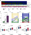
Figure 1. The scheme of AI-powered TME analyzer and the workflow of the current study. (A) Study flow diagram. (B) Landscape of AI-IP analysis. (C) Representative images of TME analyzer. (D) Alluvial plot presenting the comparison of the AI-IP and RNA expression-based TME subtype. (E) Proportional bar plot showing PD-L1 expression tumor proportion scores, according to immune phenotype. ABCP, atezolizumab plus bevacizumab, paclitaxel, and carboplatin; AI, artificial intelligence; AI-IP, artificial intelligence-powered immune-phenotype; CA, cancer area; CP, carboplatin plus paclitaxel; CS, cancer stroma; EGFR, epidermal growth factor receptor; IDP, immune-desert immune phenotype; IEP, immune-excluded immune phenotype; IIP, inflamed immune phenotype; PD-L1, programmed death-ligand 1; TIL, tumor-infiltrating lymphocyte; TKI, tyrosine kinase inhibitor; TME, tumor microenvironment; TPS, tumor proportion score.
Figure 2
Figure 2. Transcriptomic characteristics of TME assessed by AI-powered spatial analysis. (A–B) Representative figure showing the spatial distribution of AI-powered inferred cells and their matched Xenium points from spatial transcriptomics data. Comparison between cell inference based on an AI-powered TME analyzer (upper panel) and gene expression data from spatial transcriptomics (lower panel; Xenium, 10x Genomics). Spatial transcriptomic data were downloaded from the 10x Genomics website (https://www.10xgenomics.com/datasets/preview-data-ffpe-human-lung-cancer-with-xenium-multimodal-cell-segmentation-1-standard). (C–F) Volcano plots showing DEGs in the spatial transcriptomic data matched with AI-predicted cell types from H&E slides for tumor cells (C), lymphocytes (D), ECs (E), and Fibs (F). A paired H&E image and Xenium spatial transcriptomic dataset from a lung adenocarcinoma case were used, focusing on a panel of 377 tumor-related and TME-related genes. The X-axis represents the change in the cell fraction with transcript detection relative to other cell types. The cell fraction was defined as the fraction of AI-predicted cell types that expressed specific genes. The Y-axis represents the −log10(p value) calculated using Fisher’s exact test. (G) Correlation heatmap of the transcriptomics-based score and AI-powered TME analysis. (H) Scatter plots showing the correlation between transcriptomics-based scores and the AI-powered TME. AI, artificial intelligence; CA, cancer area; CAF, cancer-associated fibroblast; CS, cancer stroma; DEG, differentially expressed gene; EC, endothelial cell; Fib, fibroblast; ICR, immunological constant of rejection; TIL, tumor-infiltrating lymphocyte; TME, tumor microenvironment; TLS, tertiary lymphoid structure; VEGF, vascular endothelial growth factor.
Figure 3
Figure 3. Dynamic change of TME after acquired resistance to EGFR-TKI. (A) Overall landscape assessed by AI-IP analysis. (B) Dynamic change of PD-L1 expression after resistance to EGFR-TKI. (C) Alluvial plot presenting the change of PD-L1 expression after resistance to EGFR TKI. (D) Alluvial plot presenting the change of AI-IPs comparing pre-EGFR-TKI and post-EGFR-TKI treatment samples. (E) Dynamic change of AI-powered TME analysis in patients after resistance to EGFR-TKI. ABCP, atezolizumab plus bevacizumab, paclitaxel, and carboplatin; AI, artificial intelligence; CA, cancer area; CP, carboplatin plus paclitaxel; CS, cancer stroma; EC, endothelial cell; EGFR, epidermal growth factor receptor; Fibs, fibroblasts; TIL, tumor-infiltrating lymphocyte; TKI, tyrosine kinase inhibitor; TLS, tertiary lymphoid structure; TPS, tumor proportion score.
Figure 4
Figure 4. Dynamic change of TME according to the EGFR mutant type. Comparison analysis of pre-TKI and post-TKI TILs on CA and ECs on CA based on exon 21 L858R (A), exon 19 deletion (B), acquired T790M-positive (C), and acquired T790M-negative (D) status. P values were calculated using the Wilcoxon rank-sum test. CA, cancer area; EC, endothelial cell; EGFR, epidermal growth factor receptor; TIL, tumor-infiltrating lymphocytes.
Figure 5
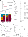
Figure 5. Efficacy outcomes with ICI after acquired resistance to EGFR-TKI. (A) Progression-free survival with ICIs monotherapy according to the IP after acquired resistance to EGFR-TKI. (B) Proportion bar plot indicating best response according to the TILs on CA, and ECs on CA. (C) Forest plot of HR (dot) and 95% CI (arrow) for PFS and OS according to the baseline characteristics and variables inferred from AI-powered TME analyzer. (D–E) Progression-free survival comparison between the ABCP arm and the PC arm in patients with higher TILs on CA (D) or ECs on CA (E) following EGFR-TKI treatment in the ATTLAS cohort. ABCP, atezolizumab plus bevacizumab, paclitaxel, and carboplatin; CA, cancer area; CS, cancer stroma; EC, endothelial cell; ECOG, Eastern Cooperative Oncology Group; EGFR, epidermal growth factor receptor; Fibs, fibroblasts; ICI, immune-checkpoint inhibitor; IP, immune-phenotype; PC, pemetrexed plus carboplatin; PD, progressive disease; PD-L1, programmed death-ligand 1; PR, partial response; SD, stable disease; TIL, tumor-infiltrating lymphocyte; TKI, tyrosine kinase inhibitor; TLS, tertiary lymphoid structure; TPS, tumor proportion score.

References

    1. Hsu W-H, Yang JC-H, Mok TS, et al. Overview of current systemic management of EGFR-mutant NSCLC. Ann Oncol. 2018;29:i3–9. doi: 10.1093/annonc/mdx702. - DOI - PubMed
    1. Ramalingam SS, Vansteenkiste J, Planchard D, et al. Overall Survival with Osimertinib in Untreated, EGFR-Mutated Advanced NSCLC. N Engl J Med. 2020;382:41–50. doi: 10.1056/NEJMoa1913662. - DOI - PubMed
    1. Cho BC, Ahn M-J, Kang JH, et al. Lazertinib Versus Gefitinib as First-Line Treatment in Patients With EGFR-Mutated Advanced Non-Small-Cell Lung Cancer: Results From LASER301. J Clin Oncol. 2023;41:4208–17. doi: 10.1200/JCO.23.00515. - DOI - PubMed
    1. Cho BC, Lu S, Felip E, et al. Amivantamab plus Lazertinib in Previously Untreated EGFR-Mutated Advanced NSCLC. N Engl J Med. 2024;391:1486–98. doi: 10.1056/NEJMoa2403614. - DOI - PubMed
    1. Planchard D, Jänne PA, Cheng Y, et al. Osimertinib with or without Chemotherapy in EGFR-Mutated Advanced NSCLC. N Engl J Med. 2023;389:1935–48. doi: 10.1056/NEJMoa2306434. - DOI - PubMed

MeSH terms

Associated data