Optimal placement of the temporary fixation pin in tibial plateau leveling osteotomy: a canine ex vivo study
- PMID: 41174761
- PMCID: PMC12577073
- DOI: 10.1186/s12917-025-05065-4
Optimal placement of the temporary fixation pin in tibial plateau leveling osteotomy: a canine ex vivo study
Abstract
Background: Tibial Plateau Leveling Osteotomy (TPLO) is widely accepted for stabilizing the stifle joint in dogs with cranial cruciate ligament disease. However, postoperative tibial tuberosity fractures remain a significant complication, particularly in small-breed dogs. Recent anatomical findings suggest that Sharpey's fibers (SF) contribute to local structural reinforcement within the tibial tuberosity, but the biomechanical impact of temporary fixation pin positioning relative to these fibers has not been experimentally quantified.
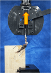
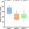
Results: Eighteen pelvic limbs from nine small-breed canine cadavers (mean body weight 5.98 kg) were randomized to three groups (n = 6) based on temporary fixation pin positioning. Group 1 had the pin inserted perpendicular to the tibial mechanical axis at the level of SF. Group 2 received pin placement 3 mm distal, and Group 3 received placement 6 mm distal and inclined from cranial to caudal. All specimens underwent a standardized TPLO, followed by mounting at a standing angle of 135°, and vertical tensile force was applied until failure. Pre- and postoperative tibial plateau angle (TPA) and absolute tibial tuberosity width (ATTW) confirmed comparable anatomy across groups. Group 1 exhibited significantly higher maximum failure loads compared to Groups 2 and 3 (p < 0.017), with no significant difference between those two groups. Fracture configuration differed notably: Group 1 showed complex, comminuted fractures of the distal tibial crest, while Groups 2 and 3 demonstrated simple linear transverse fractures through pin tract at the mid-crest.
Conclusions: Positioning the temporary fixation pin at the level of the SF markedly enhances the biomechanical resistance of the tibial tuberosity under tensile loading in ex vivo TPLO models. These findings endorse precise proximal pin placement as a modifiable surgical parameter to mitigate fracture risk in small-breed dogs. Future investigations employing dynamic loading protocols and evaluating breed-specific anatomical variations are warranted to validate these results in vivo.
Keywords: Sharpey’s fibers; Small breed dogs; Temporary fixation pin; Tensile test; Tibial plateau leveling osteotomy; Tibial tuberosity fracture.
© 2025. The Author(s).
Conflict of interest statement
Declarations. Ethics approval and consent to participate: This study utilized cadavers from ownerless dogs, donated under a formal agreement by a registered animal shelter (Incheon Veterinary Medical Association), following euthanasia for reasons unrelated to this research. As the animals were ownerless, client consent was not applicable. According to of the Konkuk University Institutional Animal Care and Use Committee (KU-IACUC) and Ministry of Agriculture, Food and Rural Affairs of South Korea, research involving cadaveric material is exempt from formal ethical review, and therefore no specific approval was required. Consent for publication: Not applicable. Competing interests: The authors declare no competing interests.
Figures


References
-
- Johnston SA, Tobias KM, editors. Veterinary surgery: small animal. Second edition. St. Louis, Missouri: Elsevier; 2018.
-
- Duval JM, Budsberg SC, Flo GL, Sammarco JL. Breed, sex, and body weight as risk factors for rupture of the cranial cruciate ligament in young dogs. J Am Vet Med Assoc. 1999;215(6):811–4. - PubMed
-
- Hayashi K, Frank JD, Dubinsky C, Hao Z, Markel MD, Manley PA, et al. Histologic changes in ruptured canine cranial cruciate ligament. Vet Surg. 2003;32(3):269–77. - PubMed
MeSH terms
LinkOut - more resources
Full Text Sources

