. 2025 Oct 1.
doi: 10.1039/d5md00706b. Online ahead of print.

Water-soluble cationic porphyrins with enhanced phototoxicity to cancer cell lines for G4-targeting photodynamic therapy

Affiliations

Water-soluble cationic porphyrins with enhanced phototoxicity to cancer cell lines for G4-targeting photodynamic therapy

Çetin Çelik et al. RSC Med Chem. .

Abstract

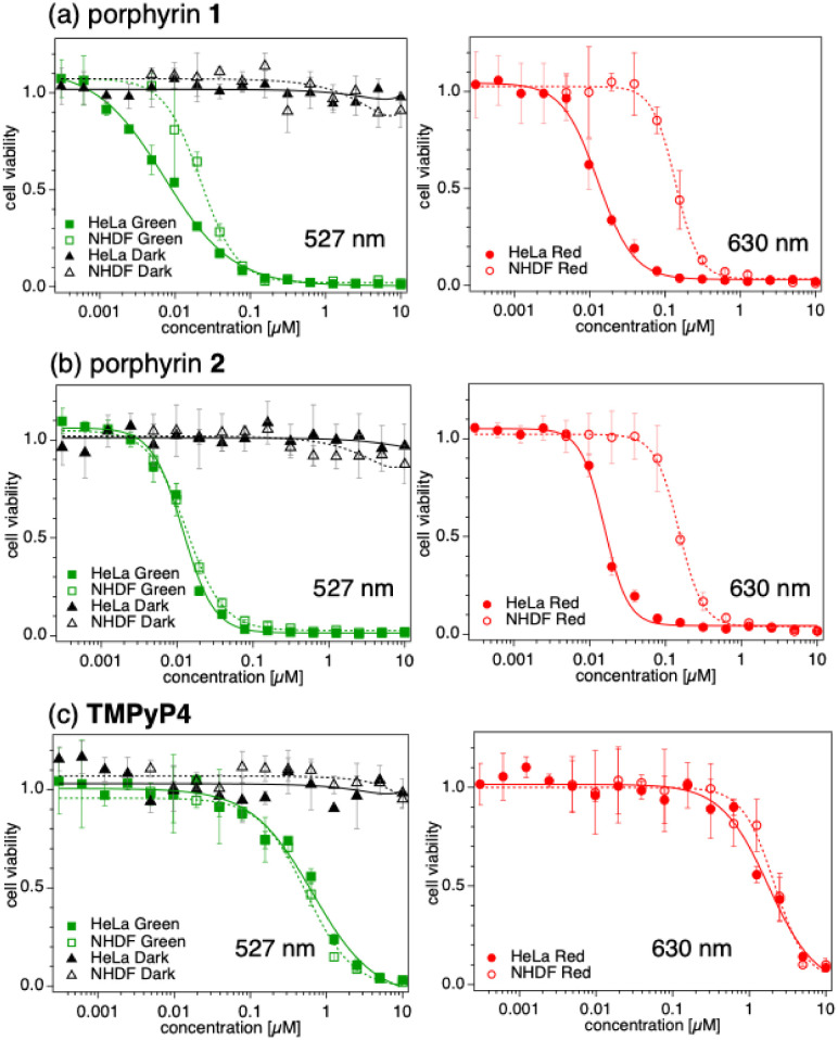
Porphyrins are well-known photosensitizers (PSs), a few of which are clinically approved drugs for use in photodynamic therapy (PDT). Porphyrin derivatives including tetra-cationic porphyrins, e.g.TMPyP4, are also well-studied binders for G-quadruplex (G4) DNA. Since G4 DNAs are known to play a role in malignant transformation of cells, a variety of G4 binders have been used in cancer therapy by regulating the function of G4 DNA. In this study, two water-soluble porphyrins (1 and 2), with four terminal cationic moieties connected with alkyl linkers were synthesized as bifunctional molecules for simultaneous G4 binding and PDT-PS. Photoinduced singlet oxygen (1O2) generation and DNA cleavage were tested under visible light irradiation revealing the efficient generation of 1O2 in line with photoinduced DNA cleavages. Studies in a cancer cell line (HeLa) and a normal fibroblast (NHDF) cells revealed significantly stronger photocytotoxicities of these porphyrins (1 and 2) in comparison to TMPyP4, presumably due to better cellular internalization - as observed by flow cytometry. Interestingly, enhanced photocytotoxicity of 1 and 2 was observed in HeLa in comparison to NHDF. This may be related to the fact that more G4 DNAs are present in the nuclei of cancer cell lines to allow binding of porphyrins 1 and 2, as observed by fluorescence microscopy. The interactions of porphyrins 1 or 2 with a G4-forming telomeric DNA were evaluated by a FRET assay and spectroscopic methods (fluorescence, UV-vis, and CD) and showed selective binding to G4 DNA. The results show the potential of porphyrins 1 and 2 as PDT-PSs targeting cancer cells with higher G4-forming domains.


Conflict of interest statement

There are no conflicts to declare.

Figures

Fig. 1
Fig. 1. Chemical structures and electrostatic surface potential maps for porphyrin 1, porphyrin 2, and TMPyP4. Conformation optimization and electrostatic surface potential calculation were performed using the universal force field operated with Avogadro 1.2.0. Blue: lower electron density; red: higher electron density.
Fig. 2
Fig. 2. UV-vis (a) and fluorescence (b) spectra of porphyrins 1, 2, and TMPyP4 (5 μM in 10 mM HEPES buffer (pH 7.4)). Fluorescence spectra were recorded with an excitation wavelength at 420 nm using a slit of 5 nm.
Fig. 3
Fig. 3. X band ESR spectra of the 1O2 adduct of 4-oxo-TEMP observed under irradiation of visible light (green LED: 539–541 nm, 90 ± 34% lm W−1; red LED: 616–626 nm, 30 ± 37% lm W−1) for 10 min. Conditions: porphyrin: 50 μM; 4-oxo-TEMP: 80 mM in pH 7.4 PBS(−).
Fig. 4
Fig. 4. (a) Photoinduced DNA cleavage of pBR322 DNA by 1, 2 and TMPyP4 under irradiation with LED light with a maximum at 527 nm (green, lanes 1–10) or at 630 nm (red, lanes 11–20) for 10 min. DNA: 12.5 μg mL−1 in Tris–HCl-EDTA buffer (pH 8.0). (b and c) Ratio of form I intact DNA after photoirradiation (b: 527 nm, 230 mW cm−2; c: 630 nm, 255 mW cm−2) in the presence of various concentrations of 1, 2, and TMPyP4, quantified using ImageJ.
Fig. 5
Fig. 5. Photocytotoxicities of porphyrins 1 (a), 2 (b) and TMPyP4 (c) under the irradiation of green LEDs (max: 527 nm max, 230 mW cm−2 for 15 min, left column) and red LEDs (max: 630 nm, 255 mW cm−2 for 15 min, right column) on HeLa and NHDF cells measured by MTT assay.
Fig. 6
Fig. 6. Flow cytometry analyses of fluorescence emission after exposure to porphyrins 1, 2 and TMPyP4 (10 μM) in HeLa (a) and NHDF (b) cell lines. Cells were incubated with porphyrins for 24 h and analysed with an excitation wavelength of 405 nm and detection of emission 692 ± 14 nm. Mean values are: (a) 399 000 (porphyrin 1), 99 500 (porphyrin 2), 26 000 (TMPyP4), and 6180 (control) and (b) 670 000 (porphyrin 1), 359 000 (porphyrin 2), 85 200 (TMPyP4), and 25 700 (control).
Fig. 7
Fig. 7. Fluorescence microscopy images of HeLa cells and NHDF cells in the presence of porphyrins 1 and 2 (5 μM). Cells were permeabilized and fixed prior to the addition of porphyrins. (Excitation: 405 nm laser, emission filter: ET700/75).
Fig. 8
Fig. 8. Fluorescence spectra of 1 (a), 2 (b) and TMPyP4 (c) (5 μM) in 10 mM HEPES, (pH 7.4 with 1 mM Na2EDTA and 100 mM KCl) in the presence of telo24 G4 DNA (0–15 μM). Excitation wavelength: 423 nm for 1, 420 nm for 2 and 432 nm for TMPyP4.
Fig. 9
Fig. 9. (a) The stabilization of G4 DNA by porphyrins, analyzed by FRET assay. The change of Tm values (ΔTm) of telo24 G4 DNA in the presence of the porphyrin 1, 2 or TMPyP4 compared to those without the compounds was plotted. (b) The selectivity for G4 DNA of porphyrins analyzed by competition FRET assay. The normalized FAM emission signals of the labeled G4 probe in the presence of competitor telo24 G4 DNA (G4) or telo24 mutant (non-G4) were shown.
Fig. 10
Fig. 10. UV-vis absorption spectra at around the Soret band of 1 (a), 2 (b) and TMPyP4 (c) (5 μM) in 10 mM HEPES (pH 7.4 with 1 mM Na2EDTA and 100 mM KCl) in the presence of telo24 G4 DNA (0–15 μM).
Fig. 11
Fig. 11. CD spectra of telo24 DNA (12.5 μM) in the presence of 0–62.5 μM of porphyrin 1 (a), porphyrin 2 (b), and TMPyP4 (c) in pH 7.4 Tris–HCl buffer (50 mM) in the presence of 100 mM KCl and 1 mM Na2EDTA.

References

    1. Lovell J. F. Liu T. W. B. Chen J. Zheng G. Chem. Rev. 2010;110:2839–2857. - PubMed
    1. Pandey R. K. and Zheng G., The Porphyrin Handbook, 2000, vol. 6, pp. 157–230
    1. Nakajima S. Hayashi H. Omote Y. Yamazaki Y. Hirata S. Maeda T. Kubo Y. Takemura T. Kakiuchi Y. Shindo Y. Koshimizu K. Sakata I. J. Photochem. Photobiol., B. 1990;7:189–198. - PubMed
    1. Woodburn K. W. Stylli S. Hill J. S. Kaye A. H. Reiss J. A. Phillips D. R. Br. J. Cancer. 1992;65:321–328. - PMC - PubMed
    1. Hiyama K. Matsui H. Tamura M. Shimokawa O. Hiyama M. Kaneko T. Nagano Y. Hyodo I. Tanaka J. Miwa Y. Ogawa T. Nakanishi T. Tamai I. J. Porphyrins Phthalocyanines. 2013;17:36–43.

LinkOut - more resources