. 2025 Oct 16:15:1654185.
doi: 10.3389/fcimb.2025.1654185. eCollection 2025.

Comprehensive profiling of host- and virus-derived circular RNAs during vesicular stomatitis virus infection

Affiliations

Comprehensive profiling of host- and virus-derived circular RNAs during vesicular stomatitis virus infection

Shanshan Miao et al. Front Cell Infect Microbiol. .

Abstract

Circular RNA (circRNA) is a new member of noncoding RNA family, which has garnered increasing attention, particularly in the context of viral infections. Vesicular stomatitis virus (VSV) is a negative-sense RNA virus that threatens animal husbandry and currently lacks effective treatments. Despite extensive studies on VSV in basic research and medical applications, the systemic profiling of circRNAs in the context of VSV remains unexplored. In this study, we conducted a comprehensive analysis of circRNA profiles in VSV-infected Vero cells using high-throughput sequencing. We identified a total of 65,645 host-derived cellular circRNAs, of which 1,682 were differentially expressed. Trend clustering revealed three significant expression patterns, and functional annotation indicated that cluster 1 was associated with proviral pathways. Subsequent results showed that VSV infection elevated the top 10 cellular circRNAs, which in turn promoted VSV replication. Additionally, we identified 120 virus-derived circRNAs, top 10 of which were upregulated by VSV and enhanced VSV infection as well. We also characterized the general features of both cellular and viral circRNAs, including genomic locations and back-splicing signals. In summary, our findings revealed that both host cellular and viral circRNAs are induced by VSV infection, subsequently affecting VSV infection. This study unveils a previously unrecognized layer of virus-host interactions involving circRNAs, which may assist in the development of control strategies for VSV and its fundamental and medical applications.

Keywords: RNA-seq2; cellular circRNAs3; differentially expressed circRNAs5; vesicular stomatitis virus1; viral circRNAs4.


Conflict of interest statement

The authors declare that the research was conducted in the absence of any commercial or financial relationships that could be construed as a potential conflict of interest.

Figures
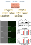
Figure 1
Figure 1
Experimental workflow and characterization of mock- and vesicular stomatitis virus (VSV)-infected Vero cells. (A) Schematic overview of the experimental workflow for identifying host- and virus-derived circular RNAs (circRNAs) in mock- and VSV-infected Vero cells. Total RNA was extracted from three conditions: Mock, 12 hours post-infection (hpi), and 24 hpi. Subsequently, RNA from each sample was ribodepleted and treated with RNase R. Library preparation was followed by sequencing on the Illumina HiSeq™ 4000 platform. Residual rRNAs were removed with Bowtie2, and the remaining reads were aligned to the Chlorocebus sabaeus genome and the VSV genome using HISAT2. Both host- and virus-derived circRNAs were identified using CIRIquant based on the detection of back-splice junction (BSJ) reads. (B) Cytopathic effects (CPE) observed in Vero cells following VSV infection. (C) Western blot analysis of VSV G protein expression. (D) Quantification of intracellular VSV RNA levels by RT-qPCR and extracellular virus titers by plaque assay. Two biological replicates were used. (A–D) All experiments were performed at a multiplicity of infection (MOI) of 0.1.
Figure 2
Figure 2
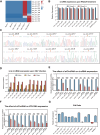
Profiling of cellular circRNAs in mock- and VSV-infected Vero cells and further differential expression analysis. (A) The number of cellular circRNAs identified within corresponding BSJ reads across all samples. (B) Genomic distribution of cellular circRNAs according to their genomic origin (including CDS, exon-intron, and intergenic), and their respective counts. (C, D) Summary of the flanking back-splice signals of cellular circRNAs. (E) Bar plot summarizing the number of differentially regulated circRNAs in each comparison groups. (F) Venn diagram illustrating the overlap and uniqueness of differentially expressed circRNAs among the indicated comparison groups. (G) Principal component analysis (PCA) based on the expression profiles of 1,682 differentially expressed cellular circRNAs. (H) Hierarchical clustering heatmap showing the expression patterns of 1,682 differentially expressed circRNAs across all samples. Each row represents a circRNA, each column represents a sample, and the values are log10-transformed reads per million (RPM).
Figure 3
Figure 3
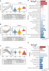
Temporal expression patterns of differentially expressed circRNAs and subsequent functional enrichment analysis. (A–C) Trend analysis of 1,682 differentially expressed circRNAs performed using Short Time-series Expression Miner (STEM). Three expression clusters were identified as statistically significant (P < 0.05). Left panels: Line plots show expression trends of circRNAs within each cluster. Gray lines represent individual circRNA expression profiles while the black line indicates the model trend for each cluster. The number of circRNAs assigned to each cluster and their corresponding P-values are provided. Right panels: Violin plots depict the absolute expression levels (log2 scale) of circRNAs within each cluster. Bottom panels: Representative circRNAs for each cluster are listed, with the corresponding source gene symbols in parentheses. (D, E) Functional enrichment analysis of the protein-coding parental genes of circRNAs from cluster 1. (D) Gene Ontology (GO) enrichment analysis results. (E) Kyoto Encyclopedia of Genes and Genomes (KEGG) pathway enrichment analysis results.
Figure 4
Figure 4
Validation of the top 10 VSV-stimulated circRNAs (VSCs) and analysis of their effects on viral replication and their subcellular localizationVero. (A) A heatmap showing the expression profiles of the top 10 VSCs, based on normalized RPM values from RNA-seq data (log10 scale). (B) RT-qPCR validation of RNase R resistance of the top 10 VSCs. Expression levels were normalized to the untreated control group. (C) Sanger sequencing of the PCR product using divergent primers to detect BSJs of the VSCs. Red arrows indicate the position of the BSJs. (D) RT-qPCR analysis of the expression levels of the top 10 VSCs in mock- and VSV-infected cells (0.1 MOI), with expression normalized to the housekeeping gene GAPDH and are presented relative to the mock-infected group. (E) RT-qPCR analysis of circRNA expression in Vero cells transfected with siRNAs targeting individual circRNAs. (F) RT-qPCR quantification of intracellular VSV RNA levels following knockdown of each VSC (excluding VSC2, VSC3, VSC5, and VSC10) by siRNA transfection, with data normalized to the negative control (siNC). *P < 0.05; **P < 0.01; ***P < 0.001; NS, no significance. (G) Subcellular localization analysis of the 10 VSCs in nuclear and cytoplasmic fractions of Vero cells using RT-qPCR, with expression levels normalized to the nuclear fraction. C, cytoplasm; N, nuclear. (B, D–G) Data are presented as mean ± SEM (n = 3).
Figure 5
Figure 5
Characterization of viral circRNAs in VSV-infected Vero cells. (A) Visualization of viral circRNAs identified in VSV-infected samples using Integrative Genomics Viewer (IGV). The viewer displays coverage the of BSJ reads mapped to the VSV genome for each infected sample, with standardized coverage depth on the Y-axis. (B) The number of viral circRNAs mapped to each VSV gene, categorized by total viral RNA (left panel), positive-sense RNA (middle panel), and negative-sense RNA (right panel). (C) Bar plots showing the total and individual counts of viral circRNAs across all infected samples. (D) Summary of the flanking signals of viral circRNAs. (E) The number of viral circRNAs detected in the corresponding BSJ reads across all samples. (F) Genomic annotation of viral circRNAs based on their origin (gene mRNA, trailer, and intergenic), along with their respective counts. (G) Distribution of viral circRNA lengths.
Figure 6
Figure 6
Expression profiling of the top 10 viral circRNAs and their impact on VSV replicationVero. (A) A heatmap illustrating the expression profiles of the top 10 VSV circRNAs (log10-transformed RPM). (B) RT-qPCR analysis of the 10 viral circRNAs following RNase R treatment. Expression levels were normalized to the untreated controls. (C) Sanger sequencing of the PCR product using divergent primers to detect BSJs of the 10 viral circRNAs, with red arrows marking the BSJ sites. (D) RT-qPCR quantification of the 10 viral circRNAs following VSV infection (0.1 MOI), with normalization to mock-infected controls. (E) RT-qPCR analysis of the expression of the 10 viral circRNAs in Vero cells transfected with siRNAs targeting each circRNA. (F) RT-qPCR quantification of VSV RNA levels upon circRNA knockdown. Data are normalized to siNC group. (G) Subcellular distribution of viral circRNAs in nuclear and cytoplasmic compartments, with the C/N ratio indicating the relative enrichment of cytoplasm over nucleus. (B, D–F) Data are presented as mean ± SEM (n = 3). *P<0.05; **P<0.01; ***P<0.001.

References

    1. Abe T., Minami N., Bawono R. G., Matsui C., Deng L., Fukuhara T., et al. (2020). ISGylation of hepatitis C virus NS5A protein promotes viral RNA replication via recruitment of cyclophilin A. J. Virol. 94, e00532-20. doi: 10.1128/jvi.00532-20, PMID: - DOI - PMC - PubMed
    1. Ahmed M. M., Okesanya O. J., Ukoaka B. M., Ibrahim A. M., Lucero-Prisno D. E., 3rd (2024). Vesicular stomatitis virus: insights into pathogenesis, immune evasion, and technological innovations in oncolytic and vaccine development. Viruses 16, 1933. doi: 10.3390/v16121933, PMID: - DOI - PMC - PubMed
    1. Barrett P. N., Mundt W., Kistner O., Howard M. K. (2009). Vero cell platform in vaccine production: moving towards cell culture-based viral vaccines. Expert Rev. Vaccines 8, 607–618. doi: 10.1586/erv.09.19, PMID: - DOI - PubMed
    1. Cai H., Holleufer A., Simonsen B., Schneider J., Lemoine A., Gad H. H., et al. (2020). 2’3’-cGAMP triggers a STING- and NF-κB-dependent broad antiviral response in Drosophila. Sci. Signal 13, eabc4537. doi: 10.1126/scisignal.abc4537, PMID: - DOI - PubMed
    1. Cai H., Li L., Slavik K. M., Huang J., Yin T., Ai X., et al. (2023). The virus-induced cyclic dinucleotide 2’3’-c-di-GMP mediates STING-dependent antiviral immunity in Drosophila. Immunity 56, 1991–2005.e1999. doi: 10.1016/j.immuni.2023.08.006, PMID: - DOI - PMC - PubMed

MeSH terms

LinkOut - more resources