. 2025 Nov 5;16(1):9761.
doi: 10.1038/s41467-025-64665-8.

Targeting SUMOylation promotes cBAF complex stabilization and disruption of the SS18::SSX transcriptome in synovial sarcoma

Affiliations

Targeting SUMOylation promotes cBAF complex stabilization and disruption of the SS18::SSX transcriptome in synovial sarcoma

Konstantinos V Floros et al. Nat Commun. .

Abstract

Synovial Sarcoma (SS) is driven by the SS18::SSX fusion oncoprotein and is ultimately refractory to therapeutic approaches. SS18::SSX alters ATP-dependent chromatin remodeling BAF (mammalian SWI/SNF) complexes, leading to the degradation of canonical (cBAF) complexes and amplified expression of SS18::SSX-containing non-canonical BAF (ncBAF or GBAF) complexes that drive an SS-specific transcription program and tumorigenesis. We demonstrate that SS18::SSX activates the SUMOylation program. The small molecule SUMOylation inhibitor, TAK-981, de-SUMOylates the cBAF/PBAF component, SMARCE1, stabilizing and restoring cBAF on chromatin, shifting SS models away from SS18::SSX-driven transcription. The result is DNA damage, cell death and tumor inhibition across both human and mouse SS tumor models. TAK-981 synergizes with cytotoxic chemotherapy through increased DNA damage, leading to tumor regression. Targeting the SUMOylation pathway in SS restores cBAF complexes and blocks the SS18::SSX transcriptome, identifying an unappreciated role of SUMOylation in SS and a subsequent therapeutic vulnerability.


Conflict of interest statement

Competing interests: A.C.F. is a consultant and equity holder in Treeline Biosciences and has previously served as a scientific advisor for AbbVie and has received research funding from IDP Pharma. K.V. receives support from AstraZeneca. R.L. is an inventor of a provisional patent on targeted killing of EBV-positive cancer cells by CRISPR/dCas9-mediated EBV reactivation. S.A.B. is Consultant for Caris Lifescience, has received honoraria from SpringWorks for an educational lecture and is senior clinical program leader at Boehringer-Ingelheim. The authors declare that these listed activities have no relationship to the present study.

Figures
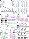
Fig. 1
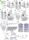
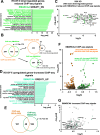
Fig. 1. Synovial sarcoma is sensitive to disruption of the SUMOylation pathway.
A Each cell type from the DepMap database was analyzed for enrichment in the custom SUMO signature (“SAE1”, “SUMO1”, “SUMO2”, “SUMO3”, “UBA2”, “UBE2I”, “RANBP2”, “CBX4”, “PIAS1”, “PIAS2”, “PIAS3”, “PIAS4”). Cell type-specific genes were ranked by the DEMETER2 v5 dependency scores (“D2_combined_gene_dep_scores.csv” file downloaded 09/18/2019) and analyzed using GSEA (clusterProfiler v.3.14.3 R package [PMID: 22455463]). The enrichment p values were −log10 transformed and used to rank the cells and plot the transformed p values on Y-axis. B RNA levels of genes involved in SUMOylation following shSSX knockdown were obtained from an RNA-seq analysis performed in SS cell lines C SS cells were transfected with siSSX for 36 h and whole cell lysates were probed with the indicated antibodies. D ChIP-seq enrichment tracks of SS18::SSX in untreated (No Rx = no drug) HS-SY-II cells compared to the tracks of the “input” at the loci of the indicated SUMO components. The combined depth-normalized signal was visualized using IGV browser and ensuring the same signal range (Y-axis) for each region of interest. E SS cells were treated with increasing concentrations of TAK-981, and cell viability was assessed 72 h later. F HS-SY-II cells underwent CRISPR/Cas9-mediated targeting of the indicated SUMOylation genes and cells were counted and plated at low density and stained with crystal violet 12 days later. G SS cells were transfected with siRNA directed against SSX for 36 h, reseeded, and treated with increasing concentrations of TAK-981 for 72 h before cell viability was assessed; inset is western blotting confirming knockdown. Protein band intensities were quantified like in (C). H SS cells were treated with 100 nM TAK-981 for 36 h or left untreated (No Rx) and whole cell lysates were probed with the indicated antibodies. H shares the same GAPDH blot with Supplementary Fig. 7C. For (E, G) n = 3 biological replicates and data are presented as mean values + SD. Unpaired two-tailed t tests were performed for comparisons between each TAK-981 treatment and No Rx for each cell line in (E) and for comparisons between siControl and siSSX in (G). Exact p values and source data can be found in the Source Data.
Fig. 2
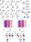
Fig. 2. TAK-981 de-SUMOylates SMARCE1, preventing its proteasomal degradation.
A Illustration depicting PTMscan SUMOylation profiling. B SUMO-2/3 complexes from untreated (No Rx) or TAK-981 treated (100 nM, 36 h) HS-SY-II cells were immunoprecipitated following transfection with exogenous SUMO2 and 36 h treatment with 100 nM of TAK-981 (or untreated cells; No Rx). Black arrow indicates SUMOylated SMARCE1. Gray arrow indicates unmodified SMARCE1 (see also Supplementary Fig. 2E). B shares the same western blots for SUMO-2/3 and TBP with Supplementary Fig. 2D. C SMARCE1 complexes were immunoprecipitated from HS-SY-II cells, following the same treatment as in (B). D Same as in (B), for Yamato cells. D shares the same western blots for SUMO-2/3 and TBP with Supplementary Fig. 2E. E Same as in (C), for Yamato cells. F HS-SY-II cells were transfected with siControl, siRNF4, siTOPORS or the siRNF4/siTOPORS, followed by treatment with 100 nM of TAK-981 for 36 h (or no treatment; No Rx) and nuclear lysates were probed with the indicated antibodies. G HS-SY-II cells from F) were analyzed by qPCR for RNF4 and TOPORS abundance. Data are values relative to the control sample and normalized to ACTB; n = 3 biological replicates; data are values + SEM. Unpaired two-tailed t tests comparing each sample with siControl. H Same as (F), for Yamato cells. I same as (G) for Yamato cells. J DepMap consortium RNAi screen data for shRNAs targeting RNF4 among 666 cancer cell lines. The RNAi score quantifies the impact of knocking down of a gene on the viability of a cell line, with more negative scores indicating a stronger dependency on the targeted gene. The boxplots indicate median and interquartile range. The whiskers extend to the minimum and to the maximum values excluding outliers. K HS-SY-II (left) and Yamato cells (right) were transfected and treated as indicated and stained with crystal violet (siR: siRNF4; siC: siControl; siT: siTOPORS). L Model of SUMO-SMARCE1 protein processing. (1) TAK-981 blocks SUMOylation and protects SMARCE1 from proteasomal degradation. (2) RNF4 tags ubiquitinated SUMO-SMARCE1. (3) In some SS cell lines, TOPORS together with RNF4 induces greater SUMO-SMARCE1 ubiquitination and proteasomal degradation. The illustrations were created in BioRender. Floros, K. (2025). Exact p values and source data can be found in the Source Data.
Fig. 3
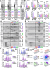
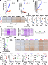
Fig. 3. SUMOylation inhibition stabilizes the cBAF complex.
A Different SS cell lines (HS-SY-II, Yamato, SYO-1 and FUJI) underwent a time-course of treatment with 100 nM of TAK-981 for 0, 12, 24, and 36 h and nuclear lysates were prepared and the expression of the indicated proteins was detected by western blotting. B The quantified signal from (A) was graphed using the GraphPad Prism software. A line was added to the No Rx levels (“1”) to simplify visual comparisons. C RNA extracts from the same cells used in (A) were analyzed by qPCR and the abundance of the mRNA levels of the indicated BAF complex subunits was quantified (n = 3 biological replicates; data are presented as mean values + SEM). Unpaired two-tailed t tests were performed for comparisons between No Rx and each time point for each BAF complex member separately. D SYO-1 cells were untreated (No Rx) or treated with 100 nM of TAK-981 for 36 h, 1 μM of TAK-243 for 3 h or 100 nM of bortezomib for 3 h and density sedimentation assay (10–30% glycerol gradient) followed by western blotting for each of the indicated BAF family components was performed on nuclear extracts. E HS-SY-II cells were infected with control shRNA (“shREN”) or shSS18::SSX (“shSSX”) and treated for 36 h with 100 nM of TAK-981, or left untreated. Density sedimentation assay (10–30% glycerol gradient) followed by the indicated BAF family components immunoblotting was performed on nuclear extracts. F Protein band intensities from D) were quantified using GeneTools software with background subtraction and normalized for each fraction to the value of No Rx for each BAF complex subunit separately (please see also Supplementary Data 2). A line was added to the No Rx levels (“1”) to simplify visual comparisons. G Protein band intensities from (E) were quantified using GeneTools software as previously (please see also Supplementary Data 3). A line was added to the shREN levels (“1”) to simplify visual comparisons. H Illustration depicting the upregulation of SMARCE1, ARID1A and BAF47 following TAK-981 treatment and the stabilization specifically of cBAF complexes (created in BioRender. Floros, K. (2025)). Exact p values and source data can be found in the Source Data.
Fig. 4
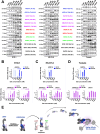
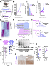
Fig. 4. TAK-981 treatment induces stronger binding of the cBAF complex to chromatin.
A Immunoblot of SS cells for specific BAF components was performed following treatment with no drug and 100 nM of TAK-981 for 36 h and differential salt extraction (0–1000 mM NaCl). GAPDH was used as a cytoplasmic loading control. Protein band intensities were quantified using GeneTools software with background subtraction and normalized to the expression of the strongest band within each blot, for drawing conclusions regarding the shift of each BAF complex component (elution at lower or higher salt concentrations) and the chromatin affinity before and after TAK-981 treatment, independent of the change in its total expression levels. B, C, D The band densities of ARID1A, SMARCE1 and BAF47 for the three SS cell lines from (A) were graphed using the GraphPad Prism software. E Illustration depicting the stabilization of the cBAF complex and the gain in its chromatin affinity after blocking SUMOylation compared to ncBAF and PBAF complexes (created in BioRender. Floros, K. (2025)). The colors that were used for the names of the BAF complex components throughout all figures of the manuscript are as follows: For unique cBAF complex subunits (e.g., ARID1A) a blue color has been used. For unique PBAF complex subunits (e.g., PBRM1 or ARID2) a green color has been used. For unique ncBAF complex subunits (e.g., BRD9 or GLTSCR1) a red color has been used. For cBAF/PBAF complex subunits (e.g., SMARCE1 or BAF47) a purple color has been used.
Fig. 5
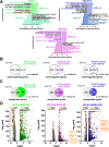
Fig. 5. Blocking SUMOylation leads to disruption of the synovial sarcoma signature and to induction of mesenchymal differentiation.
A Normalized enrichment scores after KEGG analysis of RNA-seq data demonstrating pathways altered by 100 nM TAK-981 treatment for 36 h in the indicated SS cells. B Venn diagram of genes significantly downregulated following SS18::SSX knockdown or treatment with 100 nM TAK-981 for 36 h. The significance of the gene set overlaps (p value) was calculated with Chi-square test under the assumption of 17,611 expressed transcripts. C Venn diagram of genes significantly upregulated following SS18::SSX knockdown or treatment with 100 nM TAK-981 for 36 h. The significance of the gene set overlaps (p value) was calculated with Chi-square test under the assumption of 17,611 expressed transcripts. D Volcano plot of the most down- or upregulated 1000 genes following TAK-981 treatment (detected by RNA-seq), plotted with the commonly downregulated (light orange dots) or the commonly upregulated genes (dark orange dots) after SS18::SSX KD, among the indicated SS cell line. The p values were adjusted using a False Discovery Rate (FDR) multiple testing correction method.
Fig. 6
Fig. 6. The binding of BAF complexes to chromatin is responsible for many of the observed expression changes of the SS transcriptome after blocking SUMOylation.
A Gene ontology analysis of genes associated with significantly reduced SMARCA4 ChIP-seq signals after TAK-981 treatment in HS-SY-II cells. B Venn diagrams overlap in significantly downregulated genes among the different noted experiments. The significance of the gene set overlap (p value) was calculated with Chi-squared test under the assumption of 17,611 expressed transcripts. C Overlap of the 100 most significantly downregulated genes following SS18::SSX knockdown by Banito et al. with significantly reduced SMARCA4 ChIP-seq signals. D Gene ontology analysis of genes associated with significantly upregulated SMARCA4 ChIP-seq signals after TAK-981 treatment in HS-SY-II cells. E Venn diagram of the overlap of significantly enhanced SS18::SSX and SMARCA4 ChIP-seq signals in HS-SY-II cells. The significance of the gene set overlap (p value) was calculated with Chi-square test under the assumption of 17,611 expressed transcripts. F Volcano plot of the reduced and increased SMARCA4 ChIP-seq signals plotted with the genes of commonly reduced (dark orange dots) or commonly enhanced (dark green dots) SS18::SSX ChIP-seq signals. G Genes related to the mesenchymal phenotype associated with increased SMARC4A ChIP-seq signals (green dots). padj = adjusted p values. For (A, C, D, F, G) the p values were adjusted using a False Discovery Rate (FDR) multiple testing correction method.
Fig. 7
Fig. 7. Addition of TAK-981 results in loss of chromatin accessibility to promoters of the SS18::SSX transcriptome.
A, B Boxplots demonstrating distribution of SYO-1 and HS-SY-II ATAC-seq peaks genome-wide after 100 nM of TAK-981 treatment (72 h) (n = 3 biological replicates for No Rx and n = 3 biological replicates for TAK-981-treated SYO-1 cells and n = 4 biological replicates for No Rx and n = 4 biological replicates for TAK-981 treated HS-SY-II cells), focusing on downregulated RNA-seq normalized genes for HS-SY-II cells transduced with SS18::SSX shRNA from Banito et al.. Differential accessibility peaks were annotated using the ChIPseeker v.1.40.0 R package and percentages of peaks associated with various gene-centric annotations were compared using unpaired two-sided t test. The boxplots show the median (central line), and the interquartile range of the data. The whiskers extend to the minimum and to the maximum values excluding outliers. C SYO-1 and HS-SY-II ATAC-seq profiles after TAK-981 treatment focusing on downregulated RNA-seq normalized genes of HS-SY-II cells transduced with SS18::SSX shRNA (the same list of downregulated genes used in (A, B), centered on TSS (transcription starting sites). For calculating the p values two-sided Wilcoxon tests were performed. D Schema of changes at the chromatin following TAK-981 treatment in SS. E, F Heatmaps of jointly clustered row-normalized gene expression (RNA-seq), and signal (ATAC-seq, ChIP-seq) of the top 1000 most upregulated genes and the associated peaks before and after TAK-981 treatment (number of replicates for each condition, n = 3). G Model of TAK-981 efficacy. TAK-981 deSUMOylates SMARCE1, leading to its accumulation. SMARCE1 upregulation leads to cBAF complex stabilization and redistribution of the BAF complexes (towards a normal cell phenotype) (D, G) were created in BioRender. Floros, K. (2025).
Fig. 8
Fig. 8. TAK-981 has activity in SS in vivo and sensitizes the cancer cells to cytotoxic chemotherapy.
A HS-SY-II tumor-bearing NSG mice were treated with 25 mg/kg or 50 mg/kg TAK-981 three consecutive days (Tuesday-Thursday) and tumors were monitored by caliper at least 3/w (n = 6 for control, 5 for 25 mg/kg and 6 for 50 mg/kg). B SYO-1 tumor-bearing NSG mice were treated with 25 mg/kg or 50 mg/kg TAK-981 3/w and tumors were monitored as before (n = 8 for control and 25 mg/kg and 9 for 50 mg/kg). C SS.PDX tumor-bearing NSG mice were treated with 7.5 mg/kg TAK-981 three consecutive days, the first and second week of the experiment. Drug administration was paused following the second round of treatment (day 10) and the mice were monitored for tumor growth for a total of 28 days (n = 6 for control, 5 for TAK-981). For (A–C), the data are presented as mean values + SEM. Unpaired two-tailed t tests were performed for the time point at which control mice were euthanized. D Representative images from IHC analysis of the indicated antibodies from experiment in (B). Scale bars = 100 μm. E H-scores of staining from (D). F Bliss Sum synergy scores obtained following treatment with the indicated drugs and concentrations were calculated as previously described. G SYO-1 tumor-bearing NSG mice were treated with TAK-981 (7.5 mg/kg) three consecutive days per week, for 4 weeks (28 days) and ifosfamide via intraperitoneal injection (30 mg/kg), for three consecutive days the first and 4th week of the experiment. The average tumor volume + SEM for each cohort is displayed (n = 7 for control, 9 for ifosfamide, 8 for TAK-981 and 8 for combination treatment). Unpaired two-tailed t tests compared control and treatment mice (ifosfamide, TAK-981, combination - blue asterisks, for the 13th day of treatment). Comparisons to ifosfamide (green asterisks) and to TAK-981 (purple asterisks) are depicted for the 24th day of treatment. H Representative images of IHC analyses of the indicated antibodies in SYO-1 tumors. Scale bars = 100 μm. I H-scores from (H). For (E, I), unpaired two-tailed t tests were performed. Exact p values can be found in the Source Data.
Fig. 9
Fig. 9. TAK-981 blocks tumor growth in a mouse model with conditional SS18::SSX expression.
A Schema of the design of the mouse experiment (created in BioRender. Floros, K. (2025)). B hSS2 mice were treated with 25 mg/kg 3/w or vehicle (control) and tumors were monitored by caliper at least 3/w (n = 5 for control and n = 9 for the TAK-981 treatment cohort) (n: number of tumors). The data were presented as mean values + SEM. Unpaired two-tailed t test was performed and the p value for the comparison between control and TAK-981-treated tumors was calculated, p = 0.0002. C Waterfall plot of data from (B). D Individual tumor growth from the experiment shown in (B, C). E Heatmap of the RNA expression profiles of the 1000 most differentiated genes following TAK-981 treatment, after RNA-seq analysis in the indicated hSS2 tumors (n = 3 for control and n = 6 for TAK-981 treatment). F Pathway analysis of RNA-seq data from hSS2 tumors. G Venn diagrams of significant gene changes caused by TAK-981 in hSS2 mice and the 100 most up- or down-regulated genes in vitro following SS18::SSX knockdown in HS-SY-II cells by Banito et al.. H Volcano plot of the 100 most down- or upregulated genes following SS18::SSX knockdown in vitro by Banito et al. plotted with the commonly downregulated genes (dark purple dots) or commonly upregulated genes (dark blue dots) after RNA-seq in the hSS2 tumors. The p values were adjusted using a False Discovery Rate (FDR) multiple testing correction method. I Representative tumors from the control and the TAK-981-treated cohort were harvested 2–3 h after the last TAK-981 administration and tumor lysates were subjected to western blot analysis and probed for the indicated proteins. J Representative images of IHC analysis of the indicated antibodies for control and TAK-981-treated tumors from the hSS2 mouse experiment. Scale bars = 100 μm. K H-scores of staining with the indicated antibodies from (J). Exact p values and source data can be found in the Source Data.
Fig. 10
Fig. 10. Graphical abstract of the study.
Summary of the main results of the study. 1 In SS models, blocking SUMOylation prevents SMARCE1 degradation, therefore stabilizing cBAF complexes on  chromatin and leading to redistribution of BAF complexes and subsequent cell death. 2a Blocking of SUMOylation leads to disruption of the SS signature that primarily contains genes that encode homeobox transcription factors related to neurogenesis and other developmental processes and rewires the SS cells towards a mesenchymal phenotype that contains genes encoding extracellular matrix (ECM) proteins and proteins related to muscle function (2b). 3a TAK-981 sensitizes SS to cytotoxic chemotherapy (ifosfamide) in vivo by inducing DNA damage. 3b TAK-981 blocks tumor growth in a mouse model with conditional SS18::SSX2 expression. RNA-seq analysis demonstrated pathways associated with DNA repair and the cell cycle were significantly downregulated in TAK-981-treated tumors. In contrast, pathways related to muscle function, and to ECM receptor interaction exhibited a marked upregulation in TAK-981 treated tumors. (created in BioRender. Floros, K. (2025)).

Update of

References

    1. Burningham, Z., Hashibe, M., Spector, L. & Schiffman, J. D. The epidemiology of sarcoma. Clin. Sarcoma Res.2, 14 (2012). - PMC - PubMed
    1. Clark, J. et al. Identification of novel genes, SYT and SSX, involved in the t(X;18)(p11.2;q11.2) translocation found in human synovial sarcoma. Nat. Genet7, 502–508 (1994). - PubMed
    1. Carmody Soni, E.E., Schlottman, S., Erkizan, H. V., Uren, A. & Toretsky, J. A. Loss of SS18-SSX1 inhibits viability and induces apoptosis in synovial sarcoma. Clin. Orthop. Relat. Res.472, 874–882 (2014). - PMC - PubMed
    1. Takenaka, S. et al. Downregulation of SS18-SSX1 expression in synovial sarcoma by small interfering RNA enhances the focal adhesion pathway and inhibits anchorage-independent growth in vitro and tumor growth in vivo. Int. J. Oncol.36, 823–831 (2010). - PubMed
    1. McBride, M. J. et al. The SS18-SSX fusion oncoprotein hijacks BAF complex targeting and function to drive synovial sarcoma. Cancer Cell33, 1128–1141.e1127 (2018). - PMC - PubMed

MeSH terms

LinkOut - more resources