. 2025 Nov 5;16(1):9765.
doi: 10.1038/s41467-025-64701-7.

Structural and functional characterization of TgGSK3, a druggable kinase in Toxoplasma gondii

Affiliations

Structural and functional characterization of TgGSK3, a druggable kinase in Toxoplasma gondii

Silvia Diaz-Martin et al. Nat Commun. .

Abstract

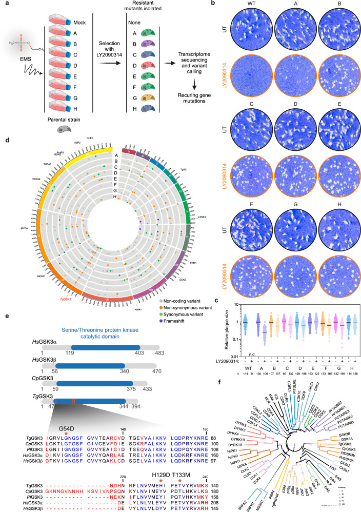
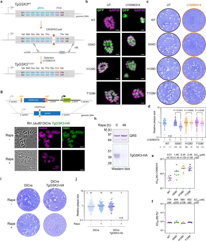
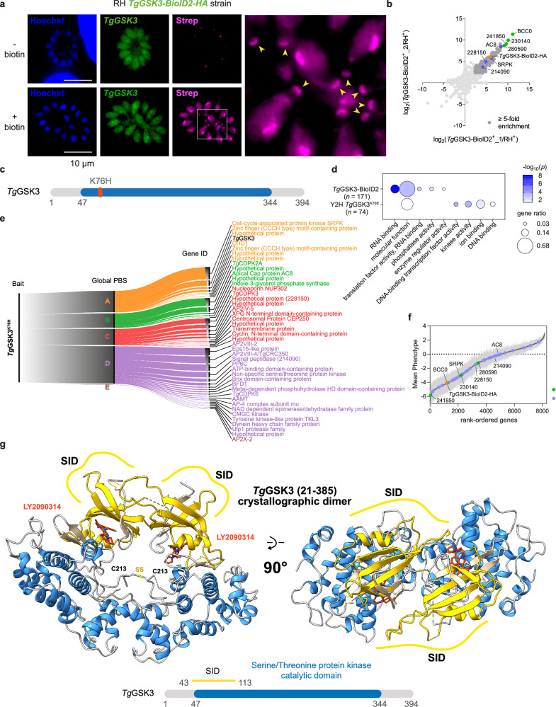
Toxoplasma gondii and Cryptosporidium species are apicomplexan parasites of significant medical and veterinary importance. Although current therapeutic options for toxoplasmosis and cryptosporidiosis demonstrate notable efficacy, their clinical efficacy is often limited by suboptimal efficacy and frequent adverse effects. Moreover, therapeutic alternatives remain limited or nonexistent, particularly for cryptosporidiosis, for which nitazoxanide is currently the only approved medication to treat diarrhea in adults and children older than 1 year of age. To identify alternative therapeutic options for addressing these health challenges, we performed a phenotypic screening of an FDA-approved drug repurposing library against Toxoplasma. This screening identifies LY2090314 as a potent inhibitor of T. gondii and Cryptosporidium growth in mammalian cells. Through a target deconvolution strategy combining forward genetics, transcriptome sequencing, and computational mutation analysis, we elucidate the parasiticidal mechanism of LY2090314 and demonstrate that TgGSK3 kinase is its primary molecular target. We also report the first X-ray crystal structure of LY2090314 bound to TgGSK3, resolved at 2.1 Å, which reveals an interaction mode characteristic of type I ATP-competitive inhibitors. Furthermore, interactome analysis uncovers functional connections between TgGSK3 and key cytoskeletal and signaling regulators, providing insights into compound's effects. Collectively, these findings validate TgGSK3 as a promising therapeutic target for toxoplasmosis and offer mechanistic insights into apicomplexan GSK3 biology.


Conflict of interest statement

Competing interests: The authors declare that they have no competing interests.

Figures

Fig. 1
Fig. 1. Effectiveness of LY2090314 against apicomplexan parasites and human host cells.
a Distribution of z-scores for each compound, calculated from the percentage of growth inhibition of T. gondii tachyzoites. The dotted line represents the cut-off mean z-score, set to select compounds with a composite z-score greater than 2, corresponding to at least 90% inhibition of parasite growth. Hoechst staining was used as a proxy for host cell viability; points are colored according to the percentage of Hoechst signal. Compounds LY2090314 (209) and the previously characterized inhibitor altiratinib (246) are highlighted. Additional GSK3 inhibitors included in the compound library are also indicated. b Chemical structure of the maleimide-based kinase inhibitor LY2090314. c Table showing the half-maximal effective concentration (EC50) values for pyrimethamine and LY2090314 against T. gondii parasites. EC50 data are presented as means ± SD from three independent biological replicates, each with three technical replicates. The half-maximal cell cytotoxicity concentration (CC50) values for LY2090314 were determined using the CellTiter-Blue or CellTox Green assay kits on HFF and ARPE-19 human cells. CC50 data are presented as means ± SD from at least two independent biological replicates, each with three technical replicates. Associated data are shown in Supplementary Fig. 1a–c. d Immunofluorescence microscopy showing the effects of LY2090314 on intracellular T. gondii parasites within HFFs. HFF cells were infected with tachyzoites (RH NLuc) and incubated with 600 nM LY2090314, 1 µM pyrimethamine (Pyr), or 0.1% DMSO as control. Cells were fixed 24 h post-infection and stained with antibodies against the T. gondii inner membrane complex protein GAP45 (magenta). Cytosolic GFP is shown in green. Scale bars, 5 µm. e Plaque assays showing the restrictive effect of LY2090314 on T. gondii lytic cycle. f The selectivity index (SI) for T. gondii parasites was calculated as the ratio of the human CC50 to the parasite EC50. g Table showing the EC50 values for LY2090314 and indirubin against C. parvum parasites. Mean EC50 values ± SD from at least three independent biological replicates are shown. Associated data are shown in Supplementary Fig. 1d, e. The CC50 data were determined using the MTS assay on HCT-8 human host cells. (n = 2). h SI for C. parvum INRAE and IOWA strains are indicated. i Evaluation of the GSK3 inhibitors BRD3731, SAR502250, and Tideglusib in plaque assays showing their lack of efficacy against T. gondii tachyzoites. j Quantification of plaque sizes shown in (i). The number of plaques analyzed is indicated above the figures. k Evaluation of BRD3731, SAR502250, and Tideglusib on C. parvum INRAE-nLuc-mCherry strain. Dose–response curves showing the percentage of host cells infected by C. parvum in response to increasing concentrations of the indicated compounds. Data were fitted using non-linear regression analysis. Results are presented as mean ± SD, with shaded error envelopes, based on at least six replicate measurements. Source data are provided as a Source Data file.
Fig. 2
Fig. 2. Target identification for LY2090314 in T. gondii parasites.
a EMS-based forward genetic strategy to isolate LY2090314-resistant T. gondii parasites. From each mutagenesis experiment, a single clone (A to H) was isolated and analyzed by RNA-seq. Created in BioRender. Hakimi, M. (https://BioRender.com/p5820hv) (b) LY2090314-resistant parasites form plaques after 7 days of growth in the presence of 600 nM LY2090314. c Quantification of plaque sizes of wild-type parasites and resistant lines (A to H) when cultured in the presence or absence of LY2090314. n.d., not detected. Data are mean from n = 3 independent experiments. d Circos plot summarizing single nucleotide variants (SNVs), insertions, and deletions detected by transcriptomic analysis of the T. gondii LY2090314-resistant lines, grouped by chromosome (numbered in Roman numerals with size intervals given outside). Each dot in the eight innermost gray tracks corresponds to a scatterplot of the mutations identified in the coding regions of the seven drug-resistant strains, with each ring representing one of the eight drug-resistant lines (A–H). Each bar in the outermost track represents locations of selected archetypal essential genes. See Supplementary Data 2 for transcriptomic analysis. e Schematic representation of the GSK3 protein architecture across different GSK3 isoforms from various species. The schematic at the top displays the conserved GSK3 catalytic domains (blue) within the GSK3 homologs from Homo sapiens Hs, Cryptosporidium parvum Cp, Plasmodium falciparum Pf, and Toxoplasma gondii Tg. Positioning of residues that were mutated in parasites resistant to LY2090314 (G54D, H129D, and T133M) are indicated in the TgGSK3 catalytic domain as orange bars. The zoomed sequence alignment below shows the amino acid sequences of the catalytic domains, with conserved residues highlighted in blue, while divergent amino acids are in red. The positions of mutations in TgGSK3 are marked with asterisks. The alignment was generated with CLC Sequence Viewer. f Phylogenetic tree of CMGC protein kinases. The tree illustrates the evolutionary relationships among various protein kinases, with branches color-coded to represent different kinase families. Bootstrap values are indicated by circle sizes. The tree was computed using the neighbor-joining algorithm implemented in Clustal Omega, with four combined iterations and the dealign input option enabled. The analysis was based on a hidden Markov model (HMM)-guided multiple sequence alignment, which included full-length sequences from the human kinome repertoire, along with GSK3 protein sequences from Cryptosporidium parvum Cp, Toxoplasma gondii Tg, Plasmodium falciparum Pf, and Plasmodium vivax Pv. The resulting tree was visualized and annotated using iTOL (Interactive Tree Of Life). Source data are provided as a Source Data file.
Fig. 3
Fig. 3. Validation of TgGSK3 as the Target of LY2090314.
a Schematic of the TgGSK3 gene editing strategy in T. gondii parasites. Detailed view of TgGSK3 locus and CRISPR/Cas9-mediated homology-directed repair with single-stranded oligo DNA nucleotides (ssODNs) carrying nucleotide substitutions (orange letters). After homologous recombination (HR) events with ssODNs, TgGSK3 recombinants were selected with LY2090314. The engineered parasites were then validated by Sanger sequencing (see Supplementary Fig. 3a). b Fluorescence microscopy showing intracellular growth of wild-type (WT, RH ku80 NLuc) and the TgGSK3-edited parasites (RH ku80 NLuc TgGSK3G54D, RH ku80 NLuc TgGSK3H129D, and RH ku80 NLuc TgGSK3T133M). HFF cells were infected with tachyzoites of the indicated T. gondii strains expressing the NLuc-P2A-EmGFP reporter gene and incubated with 600 nM LY2090314 or 0.1% DMSO as control. Cells were fixed 24 h post-infection and then stained with antibodies against the T. gondii inner membrane complex protein GAP45 (magenta). The cytosolic EmGFP is shown in green. Scale bars, 5 µm. c Effects of TgGSK3 mutations on T. gondii lytic cycle as assessed by plaque assay. Plaque sizes were measured after 7 days of growth in the presence or absence of 600 nM LY2090314. d Quantification of plaque sizes of wild-type (WT) and TgGSK3-edited parasites cultured with and without LY2090314. n.d., not detected. p values corresponding to Kruskal-Wallis test with Dunn’s multiple comparisons with the untreated (without LY2090314) conditions are indicated. Data are mean from n = 3 independent experiments. e, f EC50 values for LY2090314 (e) and pyrimethamine (Pyr) (f) were determined for WT and the TgGSK3-edited strains (G54D, H129D, and T133M). Data represent the mean from n = 6 and n = 5 independent biological replicates, each performed with three technical replicates, respectively. Mean EC50 values ± SD are indicated. g Schematic representation of the DiCre/loxP-based inducible knock-out strategy used for the conditional depletion of TgGSK3. The endogenous TgGSK3 locus of recombinant parasites (RH DiCre TgGSK3) is flanked by loxP sites (orange) and tagged with HA (purple) at the C-terminus. rec, recodonized. Below, fluorescence microscopy of the RH DiCre TgGSK3 strain after 48 h of treatment with DMSO or 50 nM rapamycin (Rapa). Cells were fixed 24 h post-infection and then stained with antibodies against GAP45 (magenta) and anti-HA antibodies (green). Scale bar, 5 µm. h Validation of Rapa-dependent depletion of TgGSK3-HA in the RH DiCre TgGSK3 strain by Western blotting analysis with anti-HA antibody. Cells were treated as in (g). TgQRS was used as loading control. i Plaque assays of the indicated strains after 7 days of growth. Cells were left untreated or treated with 50 nM Rapa. j Quantification of plaque area of n = 3 independent infections, calculated from at least 50 plaques and normalized to the mean plaque area of the parental strain. n.d., not detected. Source data are provided as a Source Data file.
Fig. 4
Fig. 4. Structural basis of LY2090314 binding and inhibition of TgGSK3.
a Overall crystallographic structure of TgGSK3 bound to LY2090314. The cartoon representation shows alpha helices in dodger blue and beta-strand in tan while the activation loop is separately colored in gold. The LY2090314 inhibitor is colored in orange and displayed in a stick representation. F196 (from the DFG motif) and phosphorylated Y211 sidechains are also displayed in a grey stick representation. N- and C-lobes are indicated. b Focus on the LY2090314 binding site. Using the same color scheme as in A, side chains involved in the compound interactions are displayed, with resistance conferring residues are shown in green while hydrogen bonds are displayed in dotted red lines and water molecules are shown as red spheres. c Rotation of the B representation by 140°. d LY2090314 binding affinity titration determined using microscale thermophoresis on wild-type and mutant TgGSK3. Normalized binding ratios are plotted against the tested LY2090314 concentrations and fitted with a Hill-Langmuir equation (used for cooperative binding models) to determine the apparent Kd (KD). WT, G54D, H129D and T133M values are plotted, respectively, in green, red, brown and blue. Data are presented as means ± SD from n = 3 independent experiments. e Endpoint kinase activity inhibition by LY2090314 on wild-type and mutant recombinant TgGSK3. WT, G54D, H129D and T133M values are plotted respectively in green, red, brown and blue. Data are presented as means ± SD from n = 3 independent experiments. Dose response curves were fitted to calculate the IC50 values. Source data are provided as a Source Data file.
Fig. 5
Fig. 5. Identification of TgGSK3-interacting proteins.
a Immunofluorescence microscopy showing TgGSK3 endogenously tagged with BioID2-HA (TgGSK3-BioID2-HA) in intracellularly growing tachyzoites. Biotinylated proteins were detected using fluorophore-conjugated streptavidin (Strep). HFF cells were infected with tachyzoites (RH TgGSK3-BioID2-HA) and incubated in the presence or absence of 150 µM biotin for 24 h prior to cell fixation and stained with antibodies against the HA tag (green), streptavidin-AlexaFluor 594 (magenta), and Hoechst (blue) to detect nuclei. Scale bars, 10 µm. b Scatter plot of the log2(ratio) of the proteins found in the two replicates (samples 1 and 2). The TgGSK3-BioID2-HA fusion protein is shown in orange, the four most highly enriched proteins are labelled in green, the candidates also identified by the yeast two-hybrid approach are indicated in blue, the proteins with at least a 5-fold enrichment in the two biological replicates are in dark grey, and the other identified proteins in light grey. c Schematic representation of the TgGSK3K76H kinase-dead mutant used as a bait in the yeast two-hybrid screening of the T. gondii cDNA library. The conserved GSK3 catalytic domains (blue) and the position of the K76E mutated residue (orange bar) are shown. d Gene ontology (GO) enrichment analysis of the proteins identified by mass spectrometry in the proximity labeling assay (TgGSK3-BioID2) and the yeast two-hybrid screening approach using TgGSK3K76E as a bait. Gene ratio is the proportion of proteins with the indicated GO term divided by the total number of proteins. Significance was determined with a hypergeometric test providing a p-value for the probability that the observed enrichment occurred by chance; only GO terms with p < 0.05 are shown. Redundant GO terms were removed. e Alluvial diagram summarizing the Yeast-two hybrid screen using TgGSK3K76H as a bait (left) and the identified partners (right) having the highest Global PBS scores. The flow diagram shows the aggregation of the hits identified into the five Global PBS score categories shown on the center. The dimension of rectangles on the right is proportional to the prevalence of phosphorylated residues for each protein (ToxoDB). f T. gondii genes rank-ordered according to their fitness scores, based on data from and ToxoDB.org. Genes identified as interactors by yeast two-hybrid (Y2H, blue) or BioID2 (green) approaches, as shown in panel (B), are highlighted. The TgGSK3-BioID2-HA fusion protein is indicated in orange. Data are presented as mean ± SEM from n = 4 independent experiments. (g) Overall crystallographic structure of disulfide linked dimer of TgGSK3 obtained from the monoclinic crystal at 3.0 Å. The cartoon representation shows alpha helices in blue and beta-strands of the N-lobes in gold while the disulfide linked between the two C213 residues in the activation loops are also displayed in a grey and gold stick representation (SS). The LY2090314 inhibitor is colored in orange and displayed in a stick representation. The Selected Interaction Domain (SID) region determined by the Y2H approach are indicated. Below, schematic representation of TgGSK3 protein architecture. Multiple independent interacting prey clones (n = 17, cell-cycle-associated protein kinase GSK, putative, Supplementary Data 4) allowed SID (in yellow) analysis that delineates the shortest fragment that is shared with all the interacting clones, and thus represents a potential region mediating the TgGSK3-TgGSK3 interaction. Source data are provided as a Source Data file.

References

    1. De Rycker, M., Baragaña, B., Duce, S. L. & Gilbert, I. H. Challenges and recent progress in drug discovery for tropical diseases. Nature559, 498–506 (2018). - PMC - PubMed
    1. World malaria report. https://www.who.int/teams/global-malaria-programme/reports/world-malaria... (2023).
    1. Djurković-Djaković, O., Dupouy-Camet, J., Van Der Giessen, J. & Dubey, J. P. Toxoplasmosis: overview from a One Health perspective. Food Waterborne Parasitol.15, e00054 (2019). - PMC - PubMed
    1. Montoya, J. G. & Liesenfeld, O. Toxoplasmosis. Lancet363, 1965–1976 (2004). - PubMed
    1. Glasner, P. D. et al. An unusually high prevalence of ocular toxoplasmosis in southern Brazil. Am. J. Ophthalmol.114, 136–144 (1992). - PubMed

MeSH terms

LinkOut - more resources