

Cytological diagnosis of ganglion cyst in a dog: A case report from a resource-limited setting
- PMID: 41200334
- PMCID: PMC12587895
- DOI: 10.5455/OVJ.2025.v15.i9.81
Cytological diagnosis of ganglion cyst in a dog: A case report from a resource-limited setting
Abstract
Background: Ganglion cysts (GCs) are rare conditions in both dogs and humans; thus, descriptions that aid in diagnosis are scarce; however, some differences exist that help us distinguish between them.
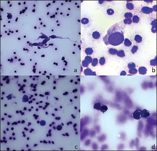
Case description: This case report presents a young female Dalmatian who developed a subcutaneous 6-cm nodule near the right ischial tuberosity, with irregular borders, slight mobility, and no pain. Fine needle aspiration cytology revealed a viscous, mucinous aspirate with sparse cellular content consistent with a GC. In this report, we discuss the most relevant findings observed under optical microscopy, present clinical data specific to this patient, and highlight the differences between ganglion and synovial cysts.
Conclusion: This case highlights the diagnostic value of fine-needle aspiration cytology, especially in settings with limited access to advanced diagnostic techniques.
Keywords: Cytology; Fine-needle aspiration; Ganglion cyst; Nodule; Synovial cyst.
Conflict of interest statement
The authors of this case report declare no conflicts of interest to declare with any person or institution.
Figures

References
-
- Aikawa T, Sadahiro S, Nishimura M, Miyazaki Y, Shibata M. Ganglion cyst arising from the composite occipito-atlanto-axial joint cavity in a cat. Vet. Comp. Orthop. Traumatol. 2014;27(4):319–23. - PubMed
-
- Christopher M.M, Hotz C.S. Cytologic diagnosis: expression of probability by clinical pathologists. Vet. Clin. Pathol. 2004;33:84–95. - PubMed
-
- Crawford A, O'Donnell M, Crowe O, Eliashar E, Smith R.K. Digital sheath synovial ganglion cysts in horses: digital sheath synovial ganglion cysts. Vet. Surg. 2011;40:66–72. - PubMed
Publication types
MeSH terms
LinkOut - more resources
Full Text Sources
Miscellaneous