. 2025 Nov;533(11):e70113.
doi: 10.1002/cne.70113.

Ophthalmologic Findings in an Induced Model of Holoprosencephaly in Zebrafish

Affiliations

Ophthalmologic Findings in an Induced Model of Holoprosencephaly in Zebrafish

Johannes Bulk et al. J Comp Neurol. 2025 Nov.

Abstract

Holoprosencephaly (HPE) is the most frequent developmental disorder of the forebrain. In this condition, the separation of the early precursor domains is hampered. A spectrum of clinical manifestations can be observed, for example, severe forms like alobar HPE and less severe forms like lobar HPE. Ophthalmologic findings that accompany HPE also occur as a spectrum that ranges from ocular hypotelorism and synophthalmia to cyclopia and anophthalmia. In this brief analysis, we made use of a recently established zebrafish model of HPE. This model is based on experimental BMP ligand induction that resulted in anophthalmia. We attenuated the induction protocol to investigate whether the ophthalmologic phenotype can also be attenuated. We found a spectrum of ocular phenotypes: ocular hypotelorism, cases of synophthalmia, and cyclopia.

Keywords: BMP; anophthalmia; bmp4; crypt‐oculoid; cyclopia; holoprosencephaly.


Conflict of interest statement

The authors declare no conflicts of interest

Figures
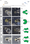
FIGURE 1
FIGURE 1
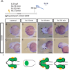
Attenuated bmp4 induction. (A) Summary of experimental procedure: embryos were heat‐shocked at 8.5 hpf for 7.5 or 10 min and analyzed at 3 dpf. (B–Dʹ) bmp4 induction for 10 min. Embryos show a spectrum of phenotypes. Frequently, two optic cups are seen that are fused in the middle, synophthalmia (B, C). However, cases of cyclopia with severe pericardial effusion (pe) can also be seen (Bʹ and Cʹ). (D and Dʹ) Schemes of phenotypes. The scheme in D matches with image C, and Dʹ matches with Cʹ. (B) Lateral view, head left. (C, Cʹ, Bʹ, D, and Dʹ) Ventral view, head left. (E–G) bmp4 induction for 7.5 min. The embryos develop ocular hypotelorism. (G) Scheme of phenotype. (E) Lateral view, head left. (F) Ventral view, head left. (H–J) Control embryo. (J) Scheme of phenotype. (H) Lateral view, head left. (I) Dorsal view, head left. Scale bars indicate 250 µm. The dotted lines indicate the eye domain; y: yolk sac. The green color in the schematic drawings indicates eye tissue of neuroectodermal origin.
FIGURE 2
FIGURE 2
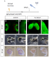
Cryaa expression in embryos after attenuated bmp4 induction. (A) Summary of experimental procedure: embryos were heat‐shocked at 8.5 hpf for 15, 10, and 7.5 min and analyzed at 48 hpf. (B–E) WMISH for cryaa at 48 hpf. (B, C) Control embryo. (B) Dorsal view, head (dotted line) left (arrow points at lenses). (C) Lateral view, head left (arrow points at lens). (D, E) bmp4‐induced embryo (15 min). (D) Dorsal view, head left. (E) Lateral view, head (dotted line) left. No eyes or lenses were formed. Multiple small cryaa‐positive domains are visible at the tip of the head (arrows in D and E). (F, G) bmp4‐induced embryo (10 min). (F) Ventral view, head left. (G) Lateral view, head left (arrows point at sites of cryaa expression). (H, I) bmp4‐induced embryo (7.5 min). (H) Ventral view, head left. (I) Lateral view, head left (arrows point at sites of cryaa expression). Scale bars indicate 200 µm. The dotted lines indicate the outlines of the heads. y: yolk sac. K: schematic representations of phenotypes. Green: eye tissue (of neuroectodermal origin); yellow: lens tissue (of ectodermal origin).
FIGURE 3
FIGURE 3
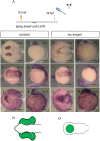
Delayed bmp4 induction results in “flat eyes.” (A) Summary of experimental procedure: embryos were heat‐shocked at 10,5 hpf (delayed) for 15 min and analyzed 24 hpf or at later stages. (B–Cʹ) Confocal images (dorsal view), with green indicating rx2‐positive cells at 24 hpf. (C, Cʹ) Control embryo. (B, Bʹ) bmp4‐induced embryo. Please note that the delayed induction resulted in out‐pocketing of optic vesicles, which, however, were not transformed into optic cups. Scale bars indicate 50 µm. (D–G) Gross analysis of embryos at 74 hpf. (D, E) Control embryo. (D) Dorsal view, head left. (E) Lateral view, head left. (F, G) bmp4‐indced embryo. (F) Dorsal view, head left. (G) Lateral view, head left. Please note that the induced embryos show a phenotype reminiscent of a flat‐eye “ojoplano” phenotype (Martinez‐Morales et al. 2009). Scale bars indicate 250 µm. (H–K) WMISH for cryaa at 48 hpf. (H, I) Control embryo. (H) Dorsal view, head left. (I) Lateral view, head left. (J, K) bmp4‐induced embryo. (J) Dorsal view, head left. (K) Lateral view, head left. Please note that the embryo shows no eyes and no visible cryaa‐positive tissue. Scale bars indicate 250 µm.
FIGURE 4
FIGURE 4
Vsx2, ofcc1, and lhx2b are expressed in crypt‐oculoids. (A) Summary of experimental procedure: embryos were heat‐shocked at 8.5 hpf for 15 min and analyzed 24 hpf. (B–E) WMISH for vsx2. (B, C) Control embryos. (B) Dorsal view, head left. (C) Lateral view, head left; vsx2 expression in optic cups. (D, E) bmp4‐induced embryos. (D) Dorsal view, head left. (E) Lateral view, head left; vsx2 expression in the head and crypt‐oculoid (arrow). (F–I) WMISH for ofcc1. (F, G) Control embryos. (F) Dorsal view, head left. (G) Lateral view, head left; ofcc1 expression in optic cups and other domains. (H, I) bmp4‐induced embryos. (D) Dorsal view, head left. (E) Lateral view, head left; ofcc1 expression in the head and crypt‐oculoid. (J–M) WMISH for lhx2b. (J, K) Control embryos. (J) Dorsal view, head left. (K) Lateral view, head left; lhx2b expression in telencephalon, diencephalon, and optic cups (arrow). (L, M) bmp4‐induced embryos. (L) Dorsal view, head left. (M) Lateral view, head left; lhx2b expression in the head and crypt‐oculoid (arrow). (N, O) Scheme of phenotype of control (N) and bmp4‐induced embryo (O); “eye tissue” is marked in green. Scale bars indicate 200 µm.
FIGURE 5
FIGURE 5
Schematic summary of findings. The eye field (green) within the ANP (center) is split and develops (green arrow) into bilateral optic cups (top), including lenses/cryaa expression domains (yellow) (arrow marks distance between optic cups). Bmp4 induction at 8.5 hpf for different durations results in graded phenotypes (7.5 min top right: ocular hypotelorism, 10 min middle right: synophthalmia/cyclopia, 15 min bottom right: crypt‐oculoid). Delayed induction of bmp4 (top left) hampers optic cup bending and results in flat eyes. Blue: forebrain.

References

    1. Addissie, Y. A. , Troia A., Wong Z. C., et al. 2021. “Identifying Environmental Risk Factors and Gene–environment Interactions in Holoprosencephaly.” Birth Defects Research 113: 63–76. - PMC - PubMed
    1. Aono, A. , Hazama M., and Notoya K.. 1995. “Potent Ectopic Bone‐Inducing Activity of Bone Morphogenetic Protein‐4/7 Heterodimer.” Biochemical and Biophysical Research Communications 210: 670–677. - PubMed
    1. Bachiller, D. , Klingensmith J., Kemp C., et al. 2000. “The Organizer Factors Chordin and Noggin Are Required for Mouse Forebrain Development.” Nature 403: 658–661. - PubMed
    1. Bogdanović, O. , Delfino‐Machín M., Nicolás‐Pérez M., et al. 2012. “Numb/Numbl‐Opo Antagonism Controls Retinal Epithelium Morphogenesis by Regulating Integrin Endocytosis.” Developmental Cell 23: 782–795. - PubMed
    1. Bulk, J. , Kyrychenko V., Rensinghoff P. M., Ghaderi Ardekani Z., and Heermann S.. 2023. “Holoprosencephaly With a Special Form of Anophthalmia Result From Experimental Induction of bmp4, Oversaturating BMP Antagonists in Zebrafish.” International Journal of Molecular Sciences 24: 8052. - PMC - PubMed

MeSH terms

Substances

LinkOut - more resources