Review
. 2025 Sep 10:22:100619.
doi: 10.1016/j.ijcchd.2025.100619. eCollection 2025 Dec.

Advances in the imaging of pulmonary hypertension

Affiliations
Review

Advances in the imaging of pulmonary hypertension

Polona Kačar et al. Int J Cardiol Congenit Heart Dis. .

Abstract

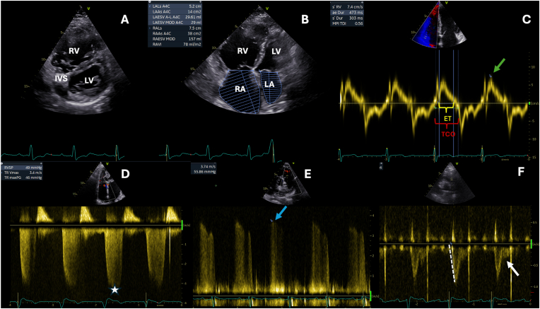
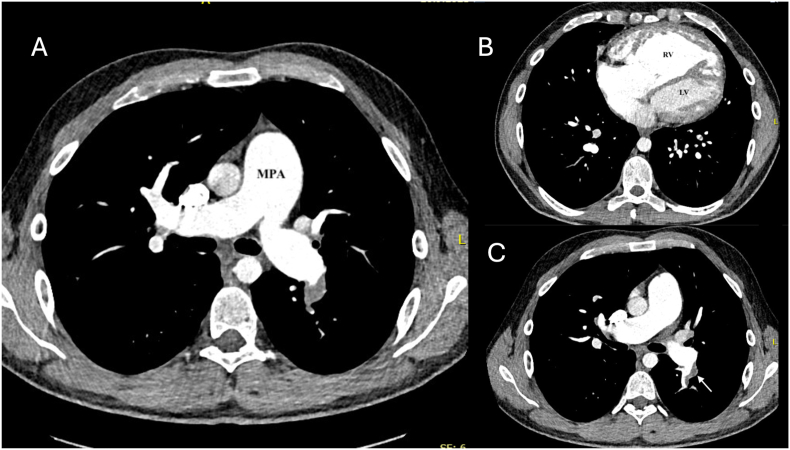
Pulmonary hypertension (PH) is a complex and progressive disorder characterized by elevated pulmonary arterial pressures leading to right ventricular dysfunction and increased morbidity and mortality. Non-invasive imaging, including echocardiography, computed tomography (CT) and cardiovascular magnetic resonance (CMR), plays a crucial role in the diagnosis, risk stratification, and management of PH. The integration of these imaging modalities facilitates a multimodal approach to PH evaluation, enabling more precise diagnosis, improved phenotyping, and better-guided therapeutic decision-making. Echocardiography remains the first-line modality, offering valuable insights into pulmonary artery pressures, right ventricular size and function, and associated cardiac anomalies. Recent developments in speckle-tracking echocardiography and 3D imaging have enhanced its diagnostic and prognostic utility. CT imaging provides detailed evaluation of the pulmonary vasculature, parenchyma, and perfusion, which is essential in distinguishing PH subtypes. CMR is non-invasive, radiation free, and highly sensitive to changes in anatomy and function making it ideal for the long-term follow up of patients with PH. It offers in-depth evaluation of all cardiac chambers as well as pulmonary blood flow assessment and tissue characterisation. In this work we discuss current strengths, limitations, and future directions in these key imaging modalities used for the comprehensive assessment of PH.


Conflict of interest statement

The authors report no relationships that could be construed as a conflict of interest other t.

Figures

Fig. 1
Fig. 1
Echocardiographic Parameters for the Assessment of Right Heart Morphology and Function A. Parasternal short axis view showing a dilated right ventricle (RV) and flattened interventricular septum (IVS) due to RV pressure overload. B. Four chamber apical view showing dilated right atrium (RA) and right ventricle (RV). C. Tissue Doppler imaging (TDI) profile of the tricuspid annulus with the measurement of systolic velocities (RV S′, green arrow) and measurement of ejection time (ET) and tricuspid closure opening time (TCO) used for calculation of the right ventricular index of myocardial performance (RIMP). D. Peak tricuspid regurgitation velocity obtained by continuous wave Doppler of the tricuspid valve (asterisk). E. Early diastolic pulmonary regurgitation velocity obtained by continuous wave Doppler of the pulmonary regurgitation (blue arrow). F. Mid-systolic notching (white arrow) and short pulmonary artery acceleration time (dashed line) of the pulmonary valve pulsed wave Doppler signal. LV, left ventricle; LA, left atrium.
Fig. 2
Fig. 2
Computed tomography pulmonary angiography (CTPA) in a patient with Eisenmenger syndrome. A. Transaxial view showing main pulmonary artery dilatation. B. Transaxial view showing enlarged right heart chambers. C. Transaxial view showing thrombus (arrow) in the interlobar portion of the left pulmonary artery.
Fig. 3
Fig. 3
Pulmonary hypertension secondary to unrepaired congenital heart disease in adults A. Pulmonary hypertension secondary to a large secundum atrial septal defect (asterisk). The right ventricle (RV) is markedly dilated and there is moderate tricuspid regurgitation. B. Unrepaired atrioventricular septal defect (asterisk) with Eisenmenger syndrome. C. Severely dilated RV with systolic flattening of the interventricular septum in keeping with RV pressure load in a patient with a large atrial septal defect. A moderate size pericardial effusion (asterisk) is present, which is a marker of poor prognosis. D. Large patent ductus arteriosus (PDA) with Eisenmenger syndrome (blue arrow). E. Unrepaired type I truncus arteriosus in a 16-year-old male with Eisenmenger syndrome. F. Large non-restrictive outlet ventricular septal defect (VSD) with Eisenmenger syndrome.
Fig. 4
Fig. 4
Tissue characterisation using cardiovascular magnetic resonance (CMR). A. A native T1 map at mid-ventricular short axis level used to determine the presence of diffuse myocardial fibrosis in the left ventricle. High T1 signal can be seen in the superior and inferior RV/LV septal insertion points (arrows). B.A similar image plane shows prominent LGE at the superior and inferior RV/LV septal insertion points. Elevated native T1 and LGE at the insertion points is common in pulmonary hypertension and likely reflects blood pooling as opposed to pathological fibrosis in pulmonary hypertension.
Fig. 5
Fig. 5
Abnormalities in pulmonary blood flow identified with cardiovascular magnetic resonance (CMR). A. Increased signal in the branch pulmonary artery and subsegmental branches indicating slow blood flow on black blood CMR images seen on HASTE (Half fourier Single-shot Turbo-spin Echo) image. B. Corresponding still from a cine image of dilated central and sub-segmental pulmonary arteries. C. Mid-systolic notch in the pulmonary arterial wave form derived from through-plane phase encoded velocity mapping. The arterial wave reflection arrowed is caused by abrupt changes in pulmonary vessel area and compliance which contribute to right ventricle afterload. This feature is indicative of pulmonary hypertension. D. In-plane 2D phase contrast image of flow showing right to left flow from the main pulmonary artery to the descending aorta (blue arrow) through the patent ductus arteriosus (blue asterisk).
Fig. 6
Fig. 6
Four-dimensional (4D) flow patterns in the pulmonary artery and quantification Image adapted from Reiter et al., 2008 (A) and Bissel et al. (2023). A. Four dimensional (4D flow map demonstrating a vortex in the forward direction of blood flow in the main pulmonary artery (MPA) in late systole. The presence of vortices in the MPA is associated with pulmonary hypertension, with the duration of the vortex existence relating to mean pulmonary artery pressures. B. Examples of position of regions of interest that can be used to quantify blood flow in the MPA, right pulmonary artery (RPA) and left pulmonary artery (LPA).
Fig. 7
Fig. 7
Three-dimensional non-contrast SSFP angiography and contrast enhance angiography demonstrating complications from PAH. Three-dimensional (3D) non-contrast dimensional balanced Steady State Free Precession (bSSFP) angiography in a patient demonstrating compression of the left main stem coronary artery between a grossly dilated pulmonary artery (PA) and aorta (Ao) as a complication of pulmonary hypertension (A and B). C. Contrast-enhanced magnetic resonance (MR) angiography in a different patient demonstrating the lumen of a grossly dilated right pulmonary artery (RPA) with thrombus in-situ. D. Still cine image of corresponding RPA with large thrombus seen proximally (white arrow) with in-situ thrombus in the left pulmonary artery (LPA) (dotted white arrow).

References

    1. Hoeper M.M., Humbert M., Souza R., Idrees M., Kawut S.M., Sliwa-Hahnle K., Jing Z.-C., Gibbs J.S.R. A global view of pulmonary hypertension. Lancet Respir Med. 2016;4:306–322. doi: 10.1016/S2213-2600(15)00543-3. - DOI - PubMed
    1. NHS Digital. National audit of pulmonary hypertension 10th annual report, Great Britain, vols. 2018–19., n.d.
    1. Mocumbi A., Humbert M., Saxena A., Jing Z.C., Sliwa K., Thienemann F., Archer S.L., Stewart S. Pulmonary hypertension. Nat Rev Dis Primers. 2024;10 doi: 10.1038/s41572-023-00486-7. - DOI - PubMed
    1. Drakopoulou M., Vlachakis P.K., Gatzoulis K., Giannakoulas G. Sudden cardiac death in pulmonary arterial hypertension. Int J Cardiol Congenit Hear Dis. 2024 doi: 10.1016/j.ijcchd.2024.100525. - DOI - PMC - PubMed
    1. Humbert M., Kovacs G., Hoeper M.M., Badagliacca R., Berger R.M.F., Brida M., Carlsen J., Coats A.J.S., Mayer E., Nagavci B., Olsson K.M., Pepke-Zaba J., Quint J.K., Simonneau G., Sitbon O., Toshner M., Delcroix M., Rosenkranz S. ESC/ERS guidelines for the diagnosis and treatment of pulmonary hypertension developed by the task force for the diagnosis and treatment of (ESC) and the european respiratory society (ERS) Eur Heart J. 2022;43:3618–3731. 2022. - PubMed

LinkOut - more resources