. 2025 Nov 11;16(1):9922.
doi: 10.1038/s41467-025-65172-6.

Persistent mirusvirus infection in the marine protist Aurantiochytrium

Affiliations

Persistent mirusvirus infection in the marine protist Aurantiochytrium

Dudley Chung et al. Nat Commun. .

Abstract

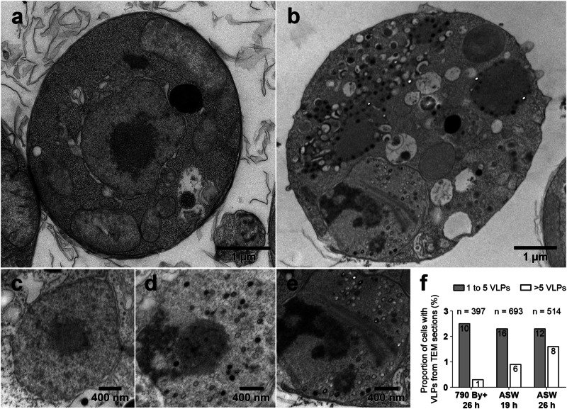
Mirusviruses are abundant and broadly distributed double-stranded (ds) DNA viruses recently discovered in marine metagenomic data. Their host range and ecological impact are unclear. The protist Aurantiochytrium limacinum possesses two mirusvirus-like genomic elements, one a circular episome (AurliV-1) and the other (AurliV-2) a chromosomal integrant. Here we show that genes in both genomes are expressed and viral particles containing mainly AurliV-1 DNA are produced under starvation conditions and when cells are cultured in standard growth medium. We detected viral particles of ~140 nm in the nucleus, in cytoplasmic vesicles, between the plasma membrane and cell wall, and in the extracellular environment. Of 67 AurliV-1-encoded proteins detected using proteomics, 45 are enriched under starvation conditions, including the structurally important major capsid and triplex proteins. Our results establish Aurantiochytrium as a model system for elucidating mirusvirus-host interactions and demonstrate persistent viral infection in a microbial eukaryote.


Conflict of interest statement

Competing interests: The authors declare no competing interests.

Figures

Fig. 1
Fig. 1. Physical maps of putative mirusvirus genomic elements of the thraustochytrid Aurantiochytrium limacinum ATCC MYA-1381.
a AurliV-1, a circular-mapping episomal mirusviral genome. b AurliV-2 is integrated into the ‘left’ end of host nuclear chromosome 15 (as designated in Collier et al. 2023) between a sub-telomeric ribosomal DNA (rDNA) operon and an internal rDNA. For AurliV-1 and V-2, all predicted ORFs ≥50 amino acids residues are shown. ORFs for which proteomic support was obtained are highlighted and mirusvirus marker genes are shown in bold. bp base pair, chr_15 chromosome 15, DNA pol DNA polymerase, DNA Top DNA topoisomerase, dUTPase deoxyuridine 5′-triphosphate nucleotidohydrolase, HJR Holliday junction resolvase, Kbp kilobase pairs, Mb megabase, MCP major capsid protein, Rpb1 RNA polymerase II subunit B1, SF2-Helicase superfamily 2 helicase, STK serine/threonine kinase, TATA-bp TATA-binding protein.
Fig. 2
Fig. 2. Morphological changes in virus-producing Aurantiochytrium limacinum cells grown under starvation conditions.
Representative TEM images of a cell lacking viral particles (a) and a virus-producing cell (b). Virus-lacking cells have a prominent central nucleolus (a, c), while virus-producing cells have peripherally located nucleoli (d, e). Fibrous structures were also observed in the nuclei of some virus-producing cells (e). f Quantification of virus-like particles (VLPs) in Aurantiochytrium limacinum cells grown under different conditions and visualized by TEM. Raw cell counts are indicated for each bar.
Fig. 3
Fig. 3. Intracellular viral particles in Aurantiochytrium limacinum.
Cell nuclei were found to contain partially and fully formed capsids (ac). Viral particles appear to acquire a coat in electron-dense regions of the cytoplasm (d, e); they appear to bud into coated vesicles (e, f), and fully formed virions accumulate between the plasma membrane and cell wall (a, f, g). Representative TEM images from at least two biological replicates are shown. Nu nucleus.
Fig. 4
Fig. 4. Extracellular viral particles from Aurantiochytrium limacinum.
Free virus particles viewed under transmission electron microscopy were found adjacent to (a) and between (b) thin electron dense layers representing shed cell wall scales and other debris in thin sectioned samples. Viral particles collected by ultracentrifugation were attached to thin layers of organic material visible by negative staining (c), which may have affected how stain penetrated the particles (df). Negative stained samples were treated with 0.5% NP-40 to expose the icosahedral capsid. Representative TEM images from at least two biological replicates are shown.
Fig. 5
Fig. 5. Droplet digital PCR analysis of mirusvirus under control and stress conditions shows AurliV-1 and AurliV-2 packaging in Aurantiochytrium limacinum.
a Absolute quantification of AurliV-1 and AurliV-2 mirusviral DNA polymerase genes, as a proxy for mirusviral particles, in the supernatant of A. limacinum cultures grown in 790 By+ (control, black circles) and artificial seawater (ASW, gray triangles). The non-mirusviral gene LeuA was quantified to measure host DNA contamination. Plots show the median (middle line), minimum and maximum values (whiskers). Three biological replicates were performed; supernatants collected at 0 h, 6 h, 18 h, 24 h and 72 h were treated with DNase during DNA extraction. b Comparison of DNase treatment on mirusvirus quantification for cultures grown in 790 By+ (black circles) and in ASW (gray triangles) media. Data from supernatants collected at 0 h, 24 h and 72 h are shown. Filled and open symbols correspond to DNase-treated (biological triplicate) and DNase-untreated (biological duplicate) samples, respectively.
Fig. 6
Fig. 6. Proteomic analysis of mirusvirus under control and stress conditions in Aurantiochytrium limacinum.
a Volcano plot of whole cell proteomic data showing differential expression of AurliV-1- and AurliV-2-derived proteins under starvation conditions (ASW) relative to cells grown in rich 790 By+ medium (control). 6101 proteins are plotted; AurliV-1-encoded proteins are in red, AurliV-2-encoded proteins in blue, and host proteins in gray. Under starvation conditions, 45 of 67 detected AurliV-1-encoded proteins were differentially expressed (i.e., log2 fold change >1 and p values adjusted for false discovery rate using a two-sided Benjamini-Hochberg test (cut-off <0.05); DA = statistically significant differentially abundant proteins; nDA = proteins not found to be differentially abundant). b Proteomic analysis of relative abundance of Aurantiochytrium limacinum AurliV-1 proteins in cells grown under control (fed) conditions (790 By+ medium) and starvation (ASW). The supernatant (i.e., extracellular) fraction under starvation conditions was also analyzed. Color coding shows annotations with major capsid protein (MCP) highlighted. Label free quantification (LFQ) was used. Viral proteins made up 0.3% to 0.7% of total annotated intensity across samples. c Predicted tertiary structure of AurliV-1 MCP generated using AlphaFold3. df Predicted hexameric structure of MCP monomer shown in (c). Colors correspond to different MCP monomers. g Select AurliV-1-encoded proteins with confident annotations detected in ASW and fed (control) samples (790 By+ medium), sorted by decreasing mean intensity from left to right. Data are presented as mean values +/− standard deviation of samples analyzed in triplicate. Individual data points are shown as open circles.

References

    1. Abergel, C., Legendre, M. & Claverie, J. M. The rapidly expanding universe of giant viruses: Mimivirus, Pandoravirus, Pithovirus and Mollivirus. FEMS Microbiol. Rev.39, 779–796 (2015). - PubMed
    1. Philippe, N. et al. Pandoraviruses: amoeba viruses with genomes up to 2.5 Mb reaching that of parasitic eukaryotes. Science341, 281–286 (2013). - PubMed
    1. Colson, P., La Scola, B., Levasseur, A., Caetano-Anolles, G. & Raoult, D. Mimivirus: leading the way in the discovery of giant viruses of amoebae. Nat. Rev. Microbiol.15, 243–254 (2017). - PMC - PubMed
    1. Aylward, F. O. et al. Taxonomic update for giant viruses in the order Imitervirales (phylum Nucleocytoviricota). Arch. Virol.168, 283 (2023). - PMC - PubMed
    1. Truchon, A. R. et al. Kratosvirus quantuckense: the history and novelty of an algal bloom disrupting virus and a model for giant virus research. Front. Microbiol.14, 1284617 (2023). - PMC - PubMed

LinkOut - more resources