. 2025 Nov 14;11(46):eadz3680.
doi: 10.1126/sciadv.adz3680. Epub 2025 Nov 12.

circHERC1-A telomerase activator

Affiliations

circHERC1-A telomerase activator

Yumeng Cui et al. Sci Adv. .

Abstract

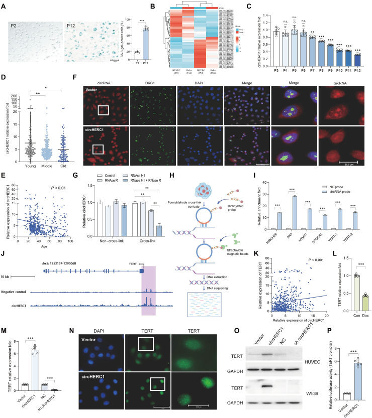
Telomerase, crucial for maintaining telomere integrity and genomic stability, is typically silenced in somatic cells with advancing age. In this study, we identify circHERC1 as a regulator of telomerase reverse transcriptase (TERT) transcription. Specifically, circHERC1 binds to the TERT promoter, facilitating the recruitment of RNA polymerase II and c-Fos, thereby activating TERT expression. Notably, circHERC1 expression exhibits a decline with age, which correlates with reduced telomerase activity. Restoration of circHERC1 expression enhances telomerase activity, promotes telomere elongation, and reverses aging-associated phenotypes. Furthermore, delivery of circHERC1 using adeno-associated virus vectors or extracellular vesicles effectively restores telomerase activity, preserves telomere integrity, and mitigates senescence. This intervention leads to improvements in cognitive function, physical performance, and a reduction in inflammation. These findings highlight the important role of circHERC1 in telomerase regulation and the aging process, positioning it as a potential therapeutic target for antiaging interventions.


Conflict of interest statement

The authors declare that they have no competing interests.

Figures

Fig. 1.
Fig. 1.. circHERC1 is associated with aging.
(A) Representative images of SA-β-gal staining of HUVECs at passage 2 (P2) and P12. (B) Heatmap of RNA sequencing (RNA-seq) data showing differential expression of circRNAs between HUVEC (P2 and P12) and HeLa (control and Dox treatment) cells. (C) qRT-PCR analysis of circHERC1 expression across cell passages in HUVECs (P3 to P12). n.s., not significant. (D) qRT-PCR analysis of circHERC1 expression in the PBMCs from healthy donors. Young (under 29 years of age), middle (from 30 to 49 years of age), and old (more than 50 years of age). (E) Correlation analysis between the expression of circHERC1 in the PBMCs from healthy donors and age. (F) Fluorescence images of circHERC1 [fluorescence in situ hybridization (FISH); red], Dyskerin 1 (DKC1) [immunofluorescence (IF); green], and 4′,6-diamidino-2-phenylindole (DAPI; blue) in circHERC1-overexpressing HeLa cells and Vector control cells. (G) qRT-PCR analysis of circHERC1 in HeLa cells after ribonuclease (RNase) R and RNase H1 digestion. (H) The diagram of circRNA-based chromatin isolation by RNA purification (CHIRP). (I) The enrichment of candidate genes by RNA pull-down with circHERC1-specific probe and negative control (NC) probe in HeLa cells. (J) CHIRP analysis indicating circHERC1 binding to genomic loci near the TERT promoter. (K) Correlation analysis between the expression of circHERC1 and TERT in the PBMCs from healthy donors. (L) qRT-PCR analysis of the expression of circHERC1 in HeLa cells treated with Dox. Con, control. (M) qRT-PCR analysis of the expression of TERT following circHERC1 overexpression or knockdown in HUVECs. (N) IF images of TERT in circHERC1-overexpressing HeLa cells and Vector control cells. (O) Western blot analysis of TERT following circHERC1 overexpression or knockdown in HUVEC (up) and WI-38 (down) cells. (P) TERT promoter luciferase assay after circHERC1 manipulation in 293FT cells. N values are shown within the panels. All values are means ± SEM. *P < 0.05, **P < 0.01, and ***P < 0.001.
Fig. 2.
Fig. 2.. circHERC1 activates TERT transcription by binding TERT promoter.
(A) Schematic diagram of truncated TERT promoter constructs used to identify regulatory regions targeted by circHERC1. The promoter regions CB1, CB3, and CB7 were highlighted as key elements in TERT transcription regulation. hTERT, human TERT. (B) Enrichment of TERT at the CB1, CB3, and CB7 promoter regions measured by qPCR after using circRNA-specific probes. (C) Msp I hypersensitivity and cross-linking assays confirmed that CB3 was an accessible and active chromatin site. (D) Ms II hypersensitivity and cross-linking assays confirmed that CB7 was an accessible and active chromatin site. (E) Site-directed mutagenesis of the CB1, CB3, and CB7 regions abolishing circHERC1-mediated transcriptional activation in 293FT cells, demonstrating that these regions are indispensable for the effect of circHERC1 on TERT promoter activity. (F) Luciferase assay assessing TERT promoter activity in response to various circHERC1 constructs in 293FT cells. (G) qRT-PCR analysis of TERT in 293FT cells transfected with circHERC1 and circHERC1 TERT binding site 1 (TB1), TB3, and TB7 mutant. (H) Western blot analysis of TERT in 293FT cells transfected with circHERC1 and circHERC1 TB1, TB3, and TB7 mutant. (I) Immunoprecipitation assay confirming an interaction between circHERC1 and RNA polymerase II in HeLa cells. pol, polymerase. (J) RNA pull-down assay demonstrated that circHERC1 bound RNA polymerase II. (K) circHERC1 mutations in polymerase II binding site 1 (PB1) and PB2 regions reduce TERT transcription. (L) The enrichment of RNA polymerase II pulled down by circHERC1-specific probe in 293FT cells transfected with circHERC1 PB mutations. (M) qRT-PCR analysis of TERT in 293FT cells transfected with circHERC1 and circHERC1 PB mutant. (N) Western blot analysis of TERT in 293FT cells transfected with circHERC1 and circHERC1 PB mutant. (O) Immunoprecipitation assay confirming an interaction between circHERC1 and c-Fos in HeLa cells. (P) RNA pull-down assay demonstrated that circHERC1 binds c-Fos in HeLa cells.
Fig. 3.
Fig. 3.. circHERC1 effectively elongates the telomere to counteract cell senescence.
(A) TRAP analysis of P3 and P12 HUVECs for telomerase activity (32 PCR circles). (B) Telomere length analysis of HUVECs at P3 to P12. (C) Telomere length–circHERC1 correlation in PBMCs of healthy donors. (D) Correlation of circHERC1 and TERT expression in PBMCs (healthy donors; CB1 site mutant). (E) Telomere length–circHERC1 correlation in PBMCs (healthy donors; CB1 site mutant). (F) TRAP analysis of HUVECs after circHERC1 overexpression and knockdown (32 PCR circles). (G) Telomere length in HUVECs after circHERC1 overexpression and knockdown. (H) Western blot of TERT in Dox-treated circHERC1-modified HeLa cells. (I) TRAP analysis of HeLa cells treated with Dox or not (28 PCR circles). (J) TRAP analysis of HeLa cells treated with H2O2 or not (28 PCR circles). (K)Telomere length of HeLa cells following circHERC1 overexpression and knockdown under conditions of DNA damage or oxidative stress. (L) TRAP analysis of 293FT cells transfected with circHERC1 and TB mutant (27 PCR circles). (M) TRAP analysis of 293FT cells transfected with circHERC1 and PB mutant (29 PCR circles). (N) Telomere length of 293FT cells transfected with circHERC1 and TB mutant. (O) Telomere length of 293FT cells transfected with circHERC1 and PB mutant. (P) Replicative life span of HUVECs with or without circHERC1 transfection. (Q) SA-β-gal staining of HUVECs after circHERC1 overexpression and knockdown. (R) Western blot of p21 and p53 in HUVECs after circHERC1 overexpression and knockdown. (S) Western blot of phospho-H2A.X in HUVECs after circHERC1 overexpression and knockdown. (T) SA-β-gal staining of HeLa cells transfected with circHERC1 and its mutants (Dox-treated). (U) Western blot of p21 and p53 in HeLa cells transfected with circHERC1 and circHERC1 PB1 and PB2 mutant. (V) Western blot of p21 and p53 in HeLa cells transfected with circHERC1 and circHERC1 TB1, TB3, and TB7 mutant. N values are shown within the panels. All values are means ± SEM.*P < 0.05, **P < 0.01, and ***P < 0.001.
Fig. 4.
Fig. 4.. circHERC1 activates TERT to preserve telomere length in vivo.
(A) qRT-PCR analysis of human circHERC1 and mouse circHERC1 in MEF cells derived from transgenic mice following tamoxifen (Tam) treatment. (B) Replicative life span of MEF cells from circHERC1 transgenic mice following Tam treatment. Tg, transgenic. (C) qRT-PCR analysis of TERT in MEF cells derived from transgenic mice following Tam treatment. (D) TRAP analysis of MEF cells derived from transgenic mice following Tam treatment (29 circles in PCR). (E) qPCR analysis of relative telomere length in MEF cells from circHERC1 transgenic mice treated with Tam. (F) Western blot analysis of TERT and p21 protein expression in young (P3) and old (P10) MEF cells derived from circHERC1 transgenic mice. (G) SA-β-gal staining in young and old MEF cells derived from circHERC1transgenic mice. (H) qRT-PCR analysis of circHERC1 expression in various tissues of circHERC1 transgenic mice. (I) Western blot analysis of TERT in various tissues of circHERC1 transgenic mice compared to controls. (J) Longitudinal analysis of telomere length in the tails of circHERC1 transgenic mice and controls. (K) qPCR analysis of relative telomere length in multiple tissues from male circHERC1 transgenic mice and controls. (L) Enzyme-linked immunosorbent assay (ELISA) analysis of serum interleukin-11 (IL-11) levels in circHERC1 transgenic mice and controls. (M) ELISA analysis of serum IL-6 levels in the serum of circHERC1 transgenic mice and controls. (N) Results from the open field test (center distance) in circHERC1 transgenic mice and controls. (O) Tail suspension test (climbing time) assessing depressive-like behavior in circHERC1 transgenic mice and controls. (P) Elevated plus maze test (open arm entry times) assessing anxiety-like behavior in circHERC1 transgenic mice and controls. N values are shown within the panels. All values are means ± SEM. *P < 0.05, **P < 0.01, and ***P < 0.001.
Fig. 5.
Fig. 5.. Effects of AAV-mediated circHERC1 overexpression in aged mice.
(A) Schematic representation of the experimental design. (B) qRT-PCR analysis of circHERC1 expression levels in different tissues from male and female mice treated with AAVcircHERC1. (C) Representative images showing the improved physical condition of male and female AAVcircHERC1-treated mice compared to controls at age of 17 months. (D) SA-β-gal staining of various tissues from AAVcircHERC1-treated male mice compared to controls. (E) Representative of SA-β-gal staining images of lung, liver, and kidney tissues from AAVcircHERC1-treated male mice compared to controls. (F) Immunohistochemical (IHC) staining for p16 and phospho-H2A.X in tissues of AAVcircHERC1-treated male mice and controls. (G) Metabolic cage experiment [respiratory exchange ratio (RER)] measuring activity levels and metabolic function in AAVcircHERC1-treated male mice and controls. (H) ELISA measurements of IL-11 in serum samples from AAVcircHERC1-treated mice and controls. (I) ELISA measurements of IL-6 in serum samples from AAVcircHERC1-treated mice and controls. (J) Open field test (velocity) between AAVcircHERC1-treated mice and controls. (K) Grip strength test (force weight) between AAVcircHERC1-treated mice and controls. (L) Western blot analysis of TERT and p21 in tissues from AAVcircHERC1-treated male mice and controls. (M) Telomere shortening rate measured in the tails of AAVcircHERC1-treated male mice and controls. (N) qRT-PCR analysis of TERT expression in various tissues from AAVcircHERC1-treated male mice and controls. (O) qPCR analysis of relative telomere length in multiple tissues from AAVcircHERC1-treated male mice and controls. N values are shown within the panels. All values are means ± SEM. *P < 0.05, **P < 0.01, and ***P < 0.001.
Fig. 6.
Fig. 6.. Evaluation of EVcirc on aging in vitro and in vivo.
(A) qRT-PCR analysis of TERT in HUVECs treated with PBS, empty EVs (EV), or EVcirc. (B) Western blot analysis of TERT and p21 expression in HUVECs treated with PBS, EV, or EVcirc. (C) TRAP analysis of HUVECs treated with PBS, EV, or EVcirc (32 circles in PCR). (D) qPCR analysis of telomere length in HUVECs treated with PBS, EV, or EVcirc. (E) SA-β-gal staining of HUVECs treated with PBS, EV, or EVcirc. (F) Western blot analysis of p21 and TERT in tissues from male mice treated with PBS, EV, or EVcirc. (G) qPCR analysis of relative telomere length in various tissues from male mice treated with PBS, EV, or EVcirc. (H) qPCR analysis of relative telomere length in various tissues from female mice treated with PBS, EV, or EVcirc. (I) SA-β-gal staining of various tissues from male mice treated with PBS, EV, or EVcirc. (J) Hematoxylin and eosin (H&E) staining of testis from male mice treated with PBS, EV, or EVcirc. (K) IHC staining of p16 and phospho-H2A.X in brains and testes of male mice treated with PBS, EV, or EVcirc. (L) MicroCT analysis of bone density in female mice treated with PBS, EV, or EVcirc. BV, bone volume; TV, total volume. (M) ELISA measurements of IL-11 in serum from mice treated with PBS, EV, or EVcirc. (N) Metabolic cage experiment (RER) in male mice treated with PBS, EV, or EVcirc. (O and P) Morris water maze test of male mice treated with PBS, EV, or EVcirc. N values are shown within the panels. All values are means ± SEM. *P < 0.05, **P < 0.01, and ***P < 0.001.

References

    1. Whittemore K., Vera E., Martínez-Nevado E., Sanpera C., Blasco M. A., Telomere shortening rate predicts species life span. Proc. Natl. Acad. Sci. U.S.A. 116, 15122–15127 (2019). - PMC - PubMed
    1. Chakravarti D., LaBella K. A., DePinho R. A., Telomeres: History, health, and hallmarks of aging. Cell 184, 306–322 (2021). - PMC - PubMed
    1. Cheng Y., Wang S., Zhang H., Lee J. S., Ni C., Guo J., Chen E., Wang S., Acharya A., Chang T. C., Buszczak M., Zhu H., Mendell J. T., A non-canonical role for a small nucleolar RNA in ribosome biogenesis and senescence. Cell 187, 4770–4789.e23 (2024). - PMC - PubMed
    1. Gorgoulis V., Adams P. D., Alimonti A., Bennett D. C., Bischof O., Bishop C., Campisi J., Collado M., Evangelou K., Ferbeyre G., Gil J., Hara E., Krizhanovsky V., Jurk D., Maier A. B., Narita M., Niedernhofer L., Passos J. F., Robbins P. D., Schmitt C. A., Sedivy J., Vougas K., von Zglinicki T., Zhou D., Serrano M., Demaria M., Cellular senescence: Defining a path forward. Cell 179, 813–827 (2019). - PubMed
    1. Miller K. N., Victorelli S. G., Salmonowicz H., Dasgupta N., Liu T., Passos J. F., Adams P. D., Cytoplasmic DNA: Sources, sensing, and role in aging and disease. Cell 184, 5506–5526 (2021). - PMC - PubMed

LinkOut - more resources