. 2025 Dec;22(12):1615-1628.
doi: 10.1038/s41423-025-01367-7. Epub 2025 Nov 12.

AIM2 drives inflammatory cell death and monkeypox pathogenesis

Affiliations

AIM2 drives inflammatory cell death and monkeypox pathogenesis

Jueun Oh et al. Cell Mol Immunol. 2025 Dec.

Abstract

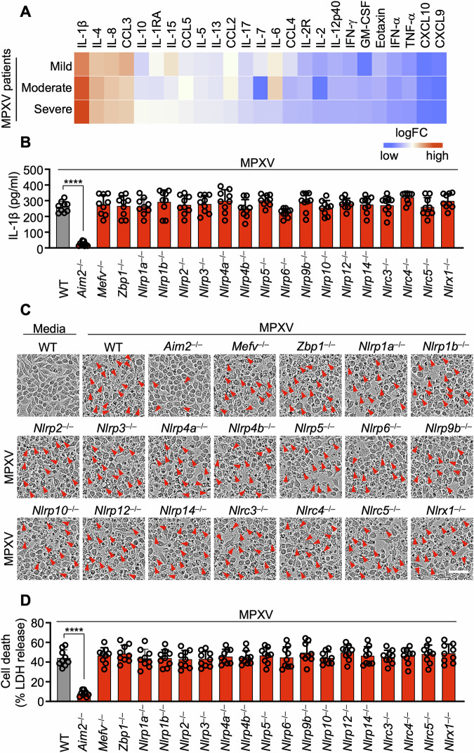
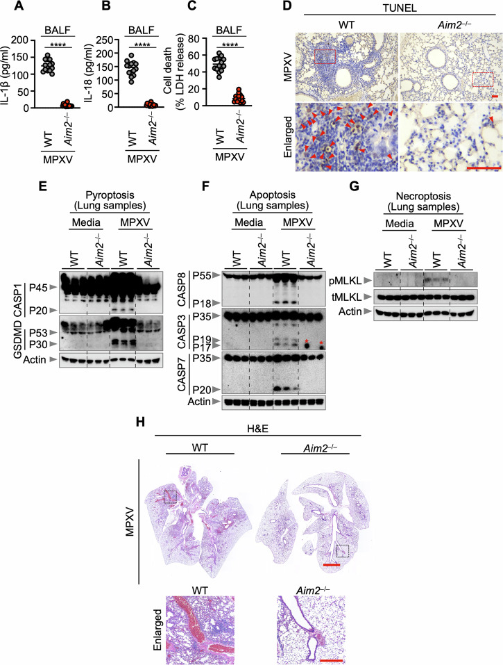
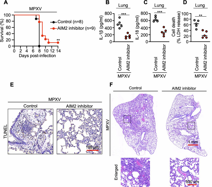
Monkeypox, a zoonotic disease caused by the monkeypox virus (MPXV), has significant global public health implications. Inflammasomes serve as crucial components of the innate immune system, detecting pathogens and triggering cell death in infected cells to eliminate harmful agents. However, the precise molecular mechanisms governing the activation of inflammasomes during MPXV infection remain largely unclear. Using CRISPR-knockout cytosolic innate immune sensor screening, we identified AIM2 as the sensor for MPXV within the inflammasome, a trigger for inflammatory cell death. Mechanistically, AIM2 forms a complex with essential cell death molecules, including ASC and caspase-1 (CASP1), without interacting with RIPK3 or CASP8. Loss of ASC, CASP1, or gasdermin D (GSDMD) reduced cell death following MPXV infection, whereas loss of GSDME, CASP3, CASP6, CASP7, CASP9, RIPK3, or MLKL did not. Pyroptotic cell death was predominantly observed in infected cells, whereas apoptotic and necroptotic signaling pathways were primarily activated in uninfected bystander cells. Furthermore, we found that the transcription factor IRF1 serves as an upstream regulator of AIM2, controlling AIM2-dependent cell death. In experiments involving AIM2-deficient mice infected with MPXV, we observed a decrease in proinflammatory cytokines, multiple inflammatory cell death pathways, and leukocyte migration, culminating in increased viral spread. CAST/EiJ mice succumbed to high-dose MPXV infection within 8 days, whereas AIM2 inhibition increased survival, with 10% of the mice treated with an AIM2 inhibitor surviving the infection. In a low-dose infection model, AIM2 inhibition reduced IL-1β and IL-18 production, LDH release, and tissue pathology. These findings highlight the critical role of AIM2-mediated inflammasome activation, along with multiple programmed cell death pathways, in shaping the innate immune response to MPXV infection, offering valuable insights for developing therapeutic strategies targeting AIM2 and the broader innate immune response against monkeypox.

Keywords: AIM2; Inflammasome; Inflammation; Inflammatory cell death; Innate immunity; Monkeypox virus.


Conflict of interest statement

Competing interests: The authors declare no competing interests.

Figures

Fig. 1
Fig. 1
Identification of AIM2 as a cytosolic innate immune sensor for monkeypox virus. A Heatmap illustrating serum cytokine/chemokine concentration levels in patients with monkeypox infection. The values for monkeypox virus (MPXV) patients were acquired from previously published cytokine assays [2], which categorize patients on the basis of disease severity. Normal values were sourced from the Bio-Plex® suspension array system tech note 6029 in Bio-Rad. The fold change was calculated for each disease severity compared with normal values as the baseline, represented on a logarithmic scale. Cytokines were ordered by the fold change under “severe” conditions. BD Assessment of IL-1β release (B), cell death (C), and lactate dehydrogenase (LDH) release (D) in wild-type (WT) or the indicated cytosolic innate immune sensor-deficient immortalized bone marrow-derived macrophages (iBMDMs) following MPXV infection (MOI of 0.1 for 24 h). B and D Data are presented as the means ± s.e.m. ****P < 0.0001 (one-way ANOVA with Dunnett’s multiple comparisons test; n = 9 from 3 biologically independent samples). (C) shows images representative of a minimum of three independent experiments. Scale bar: 50 μm
Fig. 2
Fig. 2
AIM2 is a pivotal cytosolic innate immune sensor that drives inflammasome activation and cell death in response to MPXV infection. A Immunoblot analysis of pro-caspase-1 (CASP1; P45) and cleaved CASP1 (P20) in monkeypox virus (MPXV)-infected or poly(dA:dT)-transfected wild-type (WT) or Aim2–/– bone marrow-derived macrophages (BMDMs). BE Assessment of IL-1β release (B) and IL-18 release (C) following MPXV infection (MOI of 0.1 for 24 h). D Cell death evaluation in BMDMs after MPXV infection. E Lactate dehydrogenase (LDH) release assessment after MPXV infection. FH Immunoblot analysis of CASP1 (F), cell death images (G), and LDH release (H) from WT or Aim2–/– mouse ear fibroblasts (MEFs) after MPXV infection (MOI of 0.1 for 24 h). IK Immunoblot analysis of CASP1 (I), cell death images (J), and LDH release (K) from WT or AIM2-knockdown human keratinocytes (HEK001) by short-interfering RNA (siRNA) after MPXV infection (MOI of 0.1 for 24 h). (A), (F), (I) represent data from at least three independent experiments. B, C, E, H, K Data are presented as the means ± s.e.m. “ns” not significant, ****P < 0.0001 (two-tailed t-test; n = 9 from 3 biologically independent samples). (D), (G), and (J) show images representative of a minimum of three independent experiments. Scale bar: 50 μm
Fig. 3
Fig. 3
AIM2 drives inflammatory cell death in response to monkeypox virus infection. AC Immunoblot analysis of pro-caspase-1 (CASP1; P45) and activated CASP1 (P20), pro-gasdermin D (GSDMD) (P53) and activated GSDMD (P30) (A); pro-CASP8 (P55) and cleaved CASP8 (P18), pro-CASP3 (P35) and cleaved CASP3 (P17/P19), pro-CASP7 (P35) and cleaved CASP7 (P20) (B); phosphorylated MLKL (pMLKL), total MLKL (tMLKL), phosphorylated RIPK3 (pRIPK3), and total RIPK3 (tRIPK3) (C) in bone marrow-derived macrophages (BMDMs) from WT or Aim2−/− BMDMs after monkeypox virus (MPXV) infection (MOI of 0.1 for 24 h). D Immunofluorescence (IF) images of WT or Aim2−/− BMDMs stained with MPXV and ASC at 24 h after MPXV infection (MOI of 0.1). Scale bars: 5 μm. Arrowheads indicate ASC specks. E Quantification of the percentage of ASC specks+MPXV+ cells among total cells. F Quantification of the percentage of cells with ASC specks+MPXV cells among total cells. G Immunofluorescence images of WT or Aim2−/− BMDMs stained with MPXV and cleaved caspase-3 at 24 h after MPXV infection (MOI of 0.1). Scale bars: 5 μm. H Quantification of the percentage of cleaved caspase-3 (CASP3)+MPXV+ cells among total cells. I Quantification of the percentage of cleaved CASP3+MPXV cells among total cells. J Immunofluorescence images of WT or Aim2−/− BMDMs stained with MPXV and pMLKL at 24 h after MPXV infection (MOI of 0.1). Scale bars: 5 μm. K Quantification of the percentage of pMLKL+MPXV+ cells among total cells. L Quantification of the percentage of pMLKL+MPXV cells among total cells. (A), (B), (C) represent data from at least three independent experiments. (D), (G), and (J) show images representative of a minimum of three independent experiments. Scale bar: 5 μm (merge), 6 μm (enlarged). E, F, H, I, K, L Data are presented as the means ± s.e.m. “ns” not significant, ****P < 0.0001 (two-tailed t-test; n = 9 from 3 biologically independent samples)
Fig. 4
Fig. 4
The CASP8-RIPK3 axis is not needed for the assembly of the AIM2 inflammasome in response to monkeypox virus infection. A Cell death assessment in WT, Ripk3–/–, or Ripk3−/−Casp8−/− immortalized bone marrow-derived macrophages (iBMDMs) after monkeypox virus (MPXV) infection (MOI of 0.1 for 24 h). BD Evaluation of lactate dehydrogenase (LDH) release (B), IL-1β release (C), and IL-18 release (D) following MPXV infection (MOI of 0.1 for 24 h). E Immunoprecipitation (IP) of WT and Aim2−/− BMDMs with either IgG control antibodies or anti-ASC antibodies after MPXV infection. F Immunofluorescence (IF) images of WT iBMDMs at 24 h after MPXV infection (MOI of 0.1). Arrowheads indicate ASC specks. G Quantification of the percentage of cells with ASC specks among total cells. H Quantification of the percentage of cells with ASC+AIM2+CASP1+ specks among ASC speck+ cells. (A), (F) show images representative of a minimum of three independent experiments. Scale bar: 50 μm (A), 5 μm (F). BD, G, H Data are presented as the means ± s.e.m. “ns” not significant, ****P < 0.0001 (one-way ANOVA with Dunnett’s multiple comparisons test; n = 9 from 3 biologically independent samples). E Data from at least three independent experiments are presented
Fig. 5
Fig. 5
The transcription factor IRF1 acts upstream of AIM2 and regulates cell death. A Immunoblot analysis of phosphorylated STAT1 (pSTAT1) and total STAT1 (tSTAT1) in wild-type (WT) and Ifnar1/ bone marrow-derived macrophages (BMDMs) over a defined time course following monkeypox virus (MPXV) infection (MOI of 0.1). B, C Heatmaps comparing relative gene expression between control (mock-treated) and MPXV-infected cells. B Average expression levels of interferon regulatory factor (IRF) family genes in MPXV-infected human keratinocytes relative to mock controls [29]. C Temporal expression patterns of IRF family genes in MPXV-infected MK2 cells [30]. D Immunoblot analysis of IRF1 expression in WT and Ifnar1/ BMDMs following MPXV infection (MOI of 0.1) over time. Quantification of cell death (E) and lactate dehydrogenase (LDH) release (F) in WT, Irf1/, and Aim2/ immortalized BMDMs (iBMDMs) after MPXV infection (MOI of 0.1, 24 h). Boxplots depicting the average expression levels of guanylate-binding proteins (GBPs), including GBP1 (G), GBP3 (H), GBP5 (I), GBP6 (J), and GBP7 (K), at multiple time points in MPXV-infected MK2 cells [30]. GBP2 and GBP4 were excluded because of the absence of corresponding probes. Boxes represent the median and interquartile range (IQR); whiskers indicate 1.5× IQR. (A) and (D) show representative data from at least three independent experiments. (E) presents representative images from a minimum of three independent experiments (scale bar, 50 μm). (F) shows data presented as the mean ± s.e.m.; “ns” not significant, ****P < 0.0001 (one-way ANOVA with Dunnett’s multiple comparisons test; n = 9 from three biologically independent samples)
Fig. 6
Fig. 6
AIM2 regulates inflammatory responses, viral spread, and inflammation in response to the monkeypox virus. AC Quantification of IL-1β release (A), IL-18 release (B), and lactate dehydrogenase (LDH) release (C) in bronchoalveolar lavage fluid (BALF) from WT (n = 14) and Aim2−/− (n = 14) mice on day 5 following monkeypox virus (MPXV) (western Africa strain) infection (5 × 104 PFU). D TUNEL staining of lung tissue from MPXV (western Africa strain)-infected WT and Aim2−/− mice at 5 days. TUNEL-positive cells are shown in red. EG Immunoblot analysis of pro-caspase-1 (CASP1) and activated CASP1 (P45 and P20, respectively), pro-gasdermin D (GSDMD) (P53) and activated GSDMD (P30) (E), pro-caspase-8 (CASP8) and cleaved caspase-8 (P55 and P18, respectively), pro-caspase-3 (CASP3) and cleaved CASP3 (P35, P19, and P17, respectively), pro-caspase-7 (CASP7) and cleaved CASP7 (P35 and P20, respectively) (F), and phosphorylated MLKL (pMLKL) and total MLKL (tMLKL) (G) in lung samples extracted from WT or Aim2−/− mice at 5 days post-MPXV (Western Africa strain) infection (5 × 104 PFU). The red asterisk signifies a nonspecific band. H Hematoxylin and eosin (H&E) staining of lung tissue from MPXV (western Africa strain)-infected WT and Aim2−/− mice (5 × 104 PFU). For (AC), each symbol represents an individual mouse. The data from two independent experiments were combined. The data are expressed as the means ± s.e.m. ****P < 0.0001 (two-tailed t-test). For (D), scale bars are 0.1 mm. Images are representative of five independent experiments. For (EG), each lane represents an independent biological replicate. For (H), scale bars are 2 mm (entire lung field) and 0.5 mm (enlarged field). Images are representative of five independent experiments
Fig. 7
Fig. 7
AIM2 inhibition reduces MPXV-induced mortality, inflammation, and tissue pathology in CAST/EiJ mice. A Survival of CAST/EiJ mice following high-dose intranasal challenge with monkeypox virus (MPXV, Central Africa strain; 1 × 105 PFU). The mice were intraperitoneally administered an AIM2 inhibitor (ODN TTAGGG sodium, 0.3 mg per dose) or vehicle control (DPBS) at 6 h post-infection and again on days 1, 2, and 3. BC Quantification of IL-1β (B) and IL-18 (C) levels in lung homogenates collected on day 3 post-infection (5 × 10⁴ PFU), as measured via ELISA. D Lactate dehydrogenase (LDH) release in lung homogenates as a marker of cell death (5 × 10⁴ PFU). E Representative images of TUNEL staining of lung tissue sections on day 3 post-infection (5 × 10⁴ PFU). F Hematoxylin and eosin (H&E) staining of lung sections showing inflammatory cell infiltration (5 × 10⁴ PFU). In (A), **P < 0.01 (log-rank [Mantel–Cox] test). In (BD), each symbol represents an individual mouse; data are pooled from two independent experiments and presented as the mean ± s.e.m. ***P < 0.001, **P < 0.01 (two-tailed t-test). (E) and (F) show representative images from five independent experiments

References

    1. Mitjà O, Ogoina D, Titanji BK, Galvan C, Muyembe JJ, Marks M, et al. Monkeypox. Lancet. 2023;401:60–74. - PMC - PubMed
    1. Johnston SC, Johnson JC, Stonier SW, Lin KL, Kisalu NK, Hensley LE, et al. Cytokine modulation correlates with severity of monkeypox disease in humans. J Clin Virol. 2015;63:42–5. - PMC - PubMed
    1. Li H, Huang QZ, Zhang H, Liu ZX, Chen XH, Ye LL, et al. The land-scape of immune response to monkeypox virus. EBioMedicine. 2023;87:104424. - PMC - PubMed
    1. Takeuchi O, Akira S. Pattern recognition receptors and inflammation. Cell. 2010;140:805–20. - PubMed
    1. Man SM, Karki R, Kanneganti TD. Molecular mechanisms and functions of pyroptosis, inflammatory caspases and inflammasomes in infectious diseases. Immunol Rev. 2017;277:61–75. - PMC - PubMed