. 2025 Oct 31;17(10):7638-7656.
doi: 10.21037/jtd-2025-616. Epub 2025 Oct 29.

Bioinformatics and experimental animal model reveal the prognostic value of immunogenic cell death-related proteins in idiopathic pulmonary fibrosis

Affiliations

Bioinformatics and experimental animal model reveal the prognostic value of immunogenic cell death-related proteins in idiopathic pulmonary fibrosis

Meng Li et al. J Thorac Dis. .

Abstract

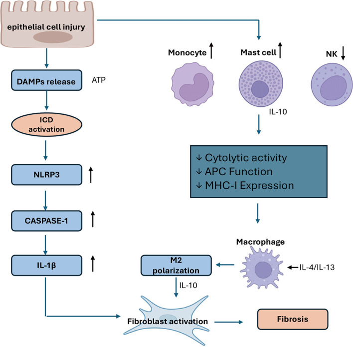
Background: Immunogenic cell death (ICD) is a type of regulated cell death (RCD) that activates adaptive immune responses and shapes the immune microenvironment. Its role in idiopathic pulmonary fibrosis (IPF), a progressive and fatal lung disease, remains unclear. This study aims to identify ICD-related gene signatures and evaluate their prognostic value in IPF through bioinformatic analysis and experimental validation.

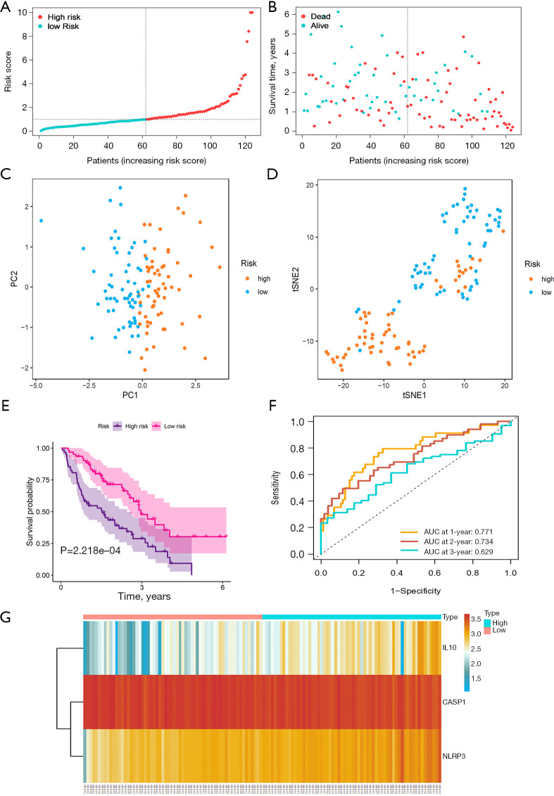
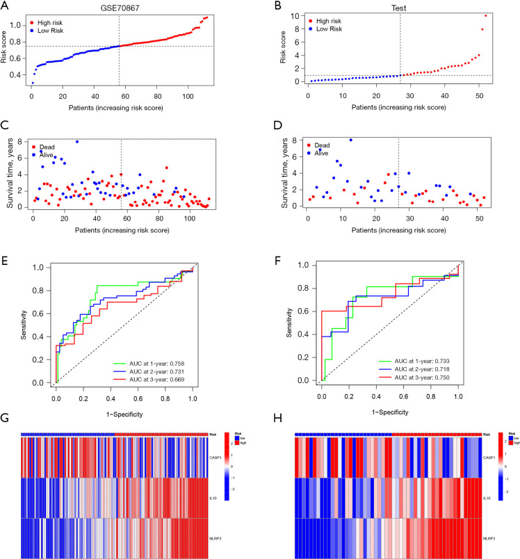
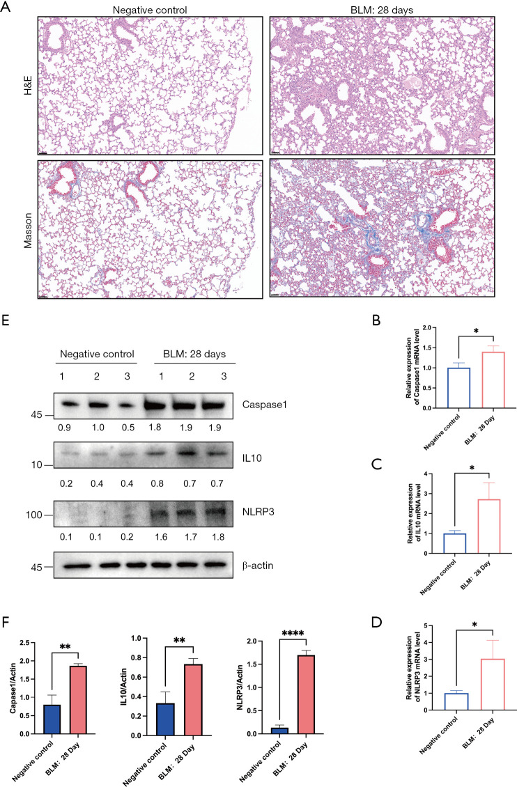
Methods: Gene expression profiles and clinical data from 176 IPF patients and 20 healthy controls were obtained from the GSE70866 dataset. A set of 34 ICD-related genes was curated from literature. Differential expression analysis, univariate Cox regression, and least absolute shrinkage and selection operator (LASSO)-penalized Cox regression were used to identify prognostic genes and construct a risk model. The model was validated internally and using an independent cohort (GSE70867). Immune cell infiltration was assessed via CIBERSORT. Expression of identified genes was further validated in a bleomycin-induced pulmonary fibrosis mouse model using quantitative real-time reverse transcription polymerase chain reaction (qRT-PCR) and Western blot.

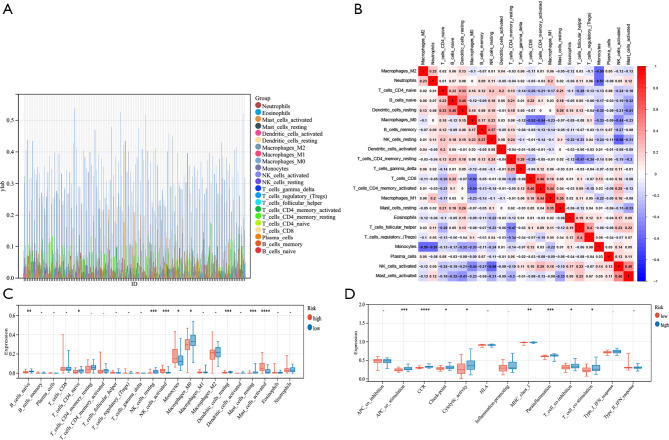
Results: Ten ICD-related genes were differentially expressed in IPF patients and associated with prognosis. A three-gene prognostic signature (IL10, CASP1, NLRP3) was established. Patients were stratified into high- and low-risk groups with significantly different overall survival (P<0.05). The risk score proved to be an independent prognostic factor for IPF. Time-dependent receiver operating characteristic (ROC) analysis showed strong predictive performance for 1-, 2-, and 3-year survival. Immune profiling revealed significant differences in mast cells, natural killer cells, and dendritic cells between risk groups. In the mouse model, mRNA and protein expression of IL10, CASP1, and NLRP3 were significantly upregulated in fibrotic lungs.

Conclusions: We developed and validated a novel ICD-related gene signature capable of predicting prognosis in IPF patients. The three-gene risk model may serve as a promising tool for risk stratification and personalized treatment planning in IPF.

Keywords: Immunogenic cell death (ICD); biomarkers; idiopathic pulmonary fibrosis (IPF); prognostic model.


Conflict of interest statement

Conflicts of Interest: All authors have completed the ICMJE uniform disclosure form (available at https://jtd.amegroups.com/article/view/10.21037/jtd-2025-616/coif). G.Z. reports that this study was supported by the Key Clinical Research Projects of The First Affiliated Hospital of Xi’an Jiaotong University (No. XJTU1AF-CRF-2023-006) and Xi’an Jiaotong University Basic-Clinical Integration Innovation Program (No. YXJLRH2022033). Y.Z. reports that this study was supported by Institutional Foundation of The First Affiliated Hospital of Xi’an Jiaotong University (No. 2024-MS-15). The other authors have no conflicts of interest to declare.

Figures

Figure 1
Figure 1
Flow chart of this study. AUC, area under the curve; BLM, bleomycin; GEO, Gene Expression Omnibus; ICD, immunogenic cell death; IPF, idiopathic pulmonary fibrosis; LASSO, least absolute shrinkage and selection operator.
Figure 2
Figure 2
Identification of prognostic ICD-related DEGs in IPF. (A) The expression levels of 12 ICD-associated DEGs in IPF patients were shown in the form of heat maps. (B) Forest plot presents the results of univariate Cox regression of the ICD-related prognosis of IPF patients. CI, confidence interval; DEGs, differentially expressed genes; ICD, immunogenic cell death; IPF, idiopathic pulmonary fibrosis.
Figure 3
Figure 3
Prognostic ICD-associated DEGs in IPF and their interrelationships. (A-C) Six ICD-associated DEGs were identified by Venn map and their expression levels were analyzed. (D,E) The correlation of six ICD-associated DEGs in total database and GSE70866 dataset. CI, confidence interval; DEGs, differentially expressed genes; ICD, immunogenic cell death; IPF, idiopathic pulmonary fibrosis.
Figure 4
Figure 4
Establishment of the ICDRS by following processes. LASSO Cox analysis identified 3 genes most associated with OS in GEO dataset (A), and the optimal λ was selected by Cross-validation (B). (C) Cox regression highlighted the correlation between ICD-regulators and IPF patients, which included 3 ICD-related genes (***, P<0.001; *, P<0.05). (D) The nomogram model for the prognosis of IPF based on CASP1, IL10, and NLRP3 gene products. (E) Calibration curve for nomogram validation. AIC, Akaike Information Criterion; GEO, Gene Expression Omnibus; ICD, immunogenic cell death; ICDRS, ICD-dependent risk profiles; IPF, idiopathic pulmonary fibrosis; LASSO, least absolute shrinkage and selection operator; OS, overall survival.
Figure 5
Figure 5
The training sets’ relationships between ICD-related genes signature and prognosis. (A,B) Risk scores distribution, overall survival were shown. (C,D) The ICDRS model effectively differentiates between the risk groups using PCA and tSNE. (E) Risk model’s prognostic significance in the training set is demonstrated by Kaplan-Meier analyses. (F) ROC curves for prognostic models based on ICDRS, predicting IPF at 1-, 2-, and 3-year intervals. (G) Heatmap of prognostic 3-gene ICDRS in training set. AUC, area under the curve; ICD, immunogenic cell death; ICDRS, ICD-dependent risk profiles; IPF, idiopathic pulmonary fibrosis; PCA, principal component analysis; ROC, receiver operating characteristic; t-SNE, t-distributed stochastic neighbor embedding.
Figure 6
Figure 6
The validation sets’ relationships between ICD-related genes signature and prognosis. (A-D) Risk scores distribution, overall survival between GSE70867 and test group. (E,F) ROC curves of 1-, 2- and 3-year prognostic models based on ICDRS for predicting IPF were identified. (G,H) Heatmaps of prognostic 3-gene ICDRS in validation set. AUC, area under the curve; ICD, immunogenic cell death; ICDRS, ICD-dependent risk profiles; IPF, idiopathic pulmonary fibrosis; ROC, receiver operating characteristic.
Figure 7
Figure 7
Univariate and multivariate Cox analyses evaluate an independent prognostic value of ICDRS in IPF patients in the training set (A,B) and the validation set (C,D). CI, confidence interval; ICD, immunogenic cell death; ICDRS, ICD-dependent risk profiles; IPF, idiopathic pulmonary fibrosis.
Figure 8
Figure 8
Bar chart of GO analysis based on the risk score-related DEGs in the training sets (A) and in the validation sets (B). Bar chart of KEGG analysis results based on the risk score-related DEGs in the training sets (C) and in the validation sets (D). BP, biological process; CC, cellular component; DEGs, differentially expressed genes; GO, Gene Ontology; KEGG, Kyoto Encyclopedia of Genes and Genomes; MF, molecular function.
Figure 9
Figure 9
The immune infiltration and function analysis in training cohort. (A) Stacking diagram by CIBERSORT algorithm. (B) Correlation matrix of ratios of immune cells. (C) Box-plot showing immune infiltration levels differ in risk groups. (D) The immune function analysis based on ssGSEA. *, P<0.05; **, P<0.01; ***, P<0.001; ****, P<0.0001. APC, antigen-presenting cell; CCR, C-C chemokine receptor; HLA, human leukocyte antigen; MHC, major histocompatibility complex; NK, natural killer; ssGSEA, single sample gene set enrichment analysis.
Figure 10
Figure 10
Verify the mRNA expression levels of the three signature genes. (A) H&E and Masson staining demonstrated that a portion of the alveolar structure was damaged in the lung of the BLM mice compared with the saline group. The qRT-PCR results of CASP1 (B), IL10 (C), and NLRP3 (D). The Western blot images results of CASP1, IL10, and NLRP3 in negative control and BLM:28 Day (E). Depression levels of CASP1, IL10, and NLRP3 in negative control and BLM:28 Day (F). The three signature genes and β-actin protein expression levels by western blotting. Scale bar: 50 μm. *, P<0.05; **, P<0.01; ****, P<0.0001. BLM, bleomycin; H&E, hematoxylin and eosin; qRT-PCR, quantitative real-time reverse transcription polymerase chain reaction.
Figure 11
Figure 11
Proposed mechanism of ICD-related proteins regulating immune microenvironment and fibrosis in IPF. In high-risk IPF patients, activation of ICD leads to the release of DAMPs, which stimulate the NLRP3-CASP1-IL1β signaling pathway. This activation is associated with increased infiltration of monocytes and mast cells, along with a reduction in NK cell levels. These immune alterations are linked to enhanced M2 macrophage polarization and elevated production of IL10 and IL13. The resulting immune environment weakens antigen presentation and cytotoxic responses, thereby facilitating fibroblast activation and progressive fibrotic remodeling. APC, antigen-presenting cell; ATP, adenosine triphosphate; DAMPs, damage-associated molecular patterns; ICD, immunogenic cell death; IPF, idiopathic pulmonary fibrosis; MHC, major histocompatibility complex; NK, natural killer.

References

    1. Richeldi L, Collard HR, Jones MG. Idiopathic pulmonary fibrosis. Lancet 2017;389:1941-52. 10.1016/S0140-6736(17)30866-8 - DOI - PubMed
    1. Lederer DJ, Martinez FJ. Idiopathic Pulmonary Fibrosis. N Engl J Med 2018;378:1811-23. 10.1056/NEJMra1705751 - DOI - PubMed
    1. Green DR, Ferguson T, Zitvogel L, et al. Immunogenic and tolerogenic cell death. Nat Rev Immunol 2009;9:353-63. 10.1038/nri2545 - DOI - PMC - PubMed
    1. Tesniere A, Panaretakis T, Kepp O, et al. Molecular characteristics of immunogenic cancer cell death. Cell Death Differ 2008;15:3-12. 10.1038/sj.cdd.4402269 - DOI - PubMed
    1. Krysko DV, Garg AD, Kaczmarek A, et al. Immunogenic cell death and DAMPs in cancer therapy. Nat Rev Cancer 2012;12:860-75. 10.1038/nrc3380 - DOI - PubMed