. 2025 Nov;55(11):e70093.
doi: 10.1002/eji.70093.

Divergent Effects of Cytomegalovirus and Rheumatoid Arthritis on Senescent CD4+ T Cells

Affiliations

Divergent Effects of Cytomegalovirus and Rheumatoid Arthritis on Senescent CD4+ T Cells

Lea Williams et al. Eur J Immunol. 2025 Nov.

Abstract

Chronic antigen exposure drives CD4⁺ T cell senescence, yet how autoimmunity and persistent viral infections differentially shape T cell differentiation and function remains unclear. Using cytomegalovirus (CMV) and rheumatoid arthritis (RA) as models of chronic immune activation, we performed high-dimensional mass cytometry and functional assays to define their impact on CD4⁺ T cells. In CMV-seropositive individuals, CD27-CD28- CD4⁺ T cells were abundant and exhibited a predominantly cytotoxic, nonproliferative phenotype. Only a minor fraction was CMV-reactive, suggesting that bystander-driven differentiation contributes to this subset. In the absence of CMV, senescent CD4⁺ T cells were infrequent and phenotypically distinct, though they still exhibited low proliferative capacity. EBV and HSV did not independently increase CD27-CD28- CD4⁺ T cell frequency. Similarly, RA had little effect on their abundance but instead tuned the functional quality of senescent cells. In CMV-seropositive RA patients, senescent CD4⁺ T cells produced less pro-inflammatory cytokines and showed impaired cytotoxic degranulation. Central memory CD4⁺ and CD27-CD28- CD8⁺ T cell functions were preserved, with no evidence for CMV reactivation, suggesting maintained viral control by unaffected T cell responses. These findings highlight distinct, nonredundant effects of CMV and RA on CD4⁺ T cell senescence and reveal RA-specific functional defects in senescent CD4⁺ T cells.


Conflict of interest statement

The authors declare no conflicts of interest.

Figures

FIGURE 1
FIGURE 1
Expansion of terminally differentiated CD4⁺ T cell subsets in CMV‐seropositive individuals. (A) UMAP displays Phenograph‐defined clusters. Data combine 5000 manually gated CD19CD3+TCRab+CD4+ cells from each donor (n = 36 donors). (B) The heatmap shows the median staining signal of individual markers for clusters shown in A. Markers used to select input cells were excluded. (C) Plot summarizes the percentage of CD4+ T cells in each cluster, divided by CMV serostatus (n = 18 per group). RM two‐way ANOVA with Sidak's multiple comparison test was performed. (D) UMAPs display the staining intensity of the indicated markers on CD4+ T cells. (E, F) Frequencies of CMV‐associated CD4⁺ T cell cluster (cluster 10) and CD8⁺ T cell clusters (clusters 5 and 7) are compared between individuals without (E) and with (F) CMV infection. Kruskal–Wallis test with Dun's multiple comparisons test was performed.
FIGURE 2
FIGURE 2
CMV‐reactive CD4⁺ T cells are enriched for the CD27CD28 phenotype. (A) Representative gating of CMV‐reactive CD4⁺ T cells. PBMCs from CMV‐seropositive individuals were stimulated with an overlapping CMV pp65 peptide pool. CMV‐reactive cells were identified by IFN‐γ and TNF‐α co‐expression. (B) Surface expression of CD27 and CD28 on TNF‐αIFN‐γ (CMV nonreactive) and TNF‐α⁺IFN‐γ⁺ (CMV‐reactive) cells. (C) Plot summarizes the frequency of CD27CD28 phenotypic subset within CMV nonreactive and CMV‐reactive populations after peptide stimulation (n = 35). Samples without detectable TNF‐α⁺IFN‐γ⁺ response were removed. Wilcoxon matched‐pairs signed rank test was performed. (D) Frequency of TNF‐α⁺IFN‐γ⁺ cells as a percentage of CD27⁺CD28⁺ or CD27CD28 CD4⁺ subsets after DMSO background subtraction. Each dot represents a response from one donor (n = 40). Wilcoxon matched‐pairs signed rank test was performed. (E) The frequency of background‐subtracted CMV‐reactive T cells as a percentage of CD27CD28 CD4+ or CD27CD28 CD8+ T cells. The Wilcoxon matched‐pairs signed rank test was performed.
FIGURE 3
FIGURE 3
CD27CD28 CD4⁺ T cells display distinct phenotypes in CMV‐seropositive and seronegative individuals. (A) GzmB, CD45RA, and CD57 co‐expression in CD27CD28 CD4+ T cells. Marker combinations were determined using Boolean operators on manually defined gates. (B) The relationship between CD27CD28 CD4+ T cell frequency and cytotoxic (GzmB+Perforin+), TEMRA (CD45RA+CD27), or CD57+ subsets. Pearson correlation was performed. (C) The frequency of GzmB+Perforin+ cells within the CD27CD28 CD4+ T cells in CMV seronegative and seropositive donors. Mann–Whitney test was performed. (D) UMAP distribution of CD27CD28 CD4+ T cells, separated by CMV status. (E) UMAPs display the staining intensity of the indicated markers. For (D) and (E), data combine comparable numbers of cells from CMV seropositive and seronegative individuals for a total of 6016 manually gated CD27CD28 CD4+ T cells. (F) Representative plot shows HLA‐DR, Ki67 staining in CD27CD28CD4+ T cells from a CMV seronegative individual. (G) Plot summarizes the frequencies of Ki67+HLA‐DR+ T cells as a percentage of CD27CD28 CD4+ subset in CMV‐seronegative or seropositive individuals. Mann–Whitney test was performed. (H) The correlation between CD27CD28 CD4+ T cell frequency and the abundance of Ki67+HLA‐DR+ cells within this subset. Each filled circle represents data from one individual (n = 36). Pearson correlation was performed. GzmB: granzyme B.
FIGURE 4
FIGURE 4
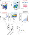
CD27CD28 CD4⁺ T cells show impaired proliferation independent of CMV status. (A) Representative plots show gating of CD27CD28 CD4+ T cells and non‐CD27CD28 memory cells (other memory). (B) CD27CD28 CD4+ T cells and other memory cells were sorted for stimulation with CD3/CD28 Dynabeads for 5 days. Plots show the percentage of the divided CTV low (CTVlo) subset. (C) The histogram shows the CTV profiles of CD27CD28 versus other memory CD4⁺ T cells. (D) Summary plot of the frequency of CTVlo cells in CD27CD28 and other CD4+ memory subsets. Each filled circle represents data from one individual (n = 10). The Wilcoxon matched‐pairs signed rank test was performed. (E) Frequency of CTVlo cells within CD27CD28 CD4+ subset after dynabead stimulation, grouped by CMV serostatus. Mann–Whitney test was used.
FIGURE 5
FIGURE 5
RA, EBV, and HSV have limited impact on senescent CD4⁺ T cell frequencies. (A) Frequencies of CD27CD28 subset as a percentage of CD4+ T cells, grouped by CMV, EBV, and HSV seropositivity. Each filled circle represents data from one individual (n = 36). Kruskal–Wallis and Dunn's multiple comparison tests were used. (B) Frequencies of GzmB and perforin‐expressing CD4+ T cells from EBV and HSV‐seropositive donors, with or without CMV co‐infection. Mann–Whitney test was performed. (C) The ratio of averaged GzmB+Perforin+ expression between CMV seropositive and seronegative individuals with EBV and HSV co‐infection, grouped by RA status. (D) UMAPs of CD27CD28CD4+ T cells, separated by CMV serostatus and RA diagnosis. CMV‐RA‐ (CMV seronegative controls), CMV‐RA+ (CMV seronegative RA patients), CMV+RA‐ (CMV seropositive controls), CMV+RA+ (CMV seropositive RA patients). (E–G) Plots summarize the percentage of CD4+ T cells in cluster 10 (E), negative for CD27 and CD28 (F), or expressing GzmB and perforin (G) by CMV and RA status. Kruskal‐Wallis and Dunn's multiple comparison tests were performed. GzmB: granzyme B.
FIGURE 6
FIGURE 6
Senescent CD4+ T cells display decreased functional potential in RA patients. (A) PBMCs from 30 CMV‐seropositive healthy controls and 29 CMV‐seropositive RA patients were stimulated with SEB for 6 h and analyzed for cytokine production and degranulation. Representative flow plots show gating for the indicated markers under vehicle control and SEB‐stimulated conditions. (B) Frequencies of TNF‐α⁺, IFN‐γ⁺, and CD107a⁺ cells within CD27CD28 CD4⁺ T cells in RA patients and controls. (C) Ratio of CD107a to CD107a⁺ cells within the CD27CD28 CD4⁺ T cell subset. (D) Frequency of polyfunctional CD27CD28 CD4⁺ T cells co‐expressing TNF‐α, IFN‐γ, and CD107a, grouped by RA status. (E) CMV‐seropositive individuals, with or without RA, were analyzed for CMV DNA by quantitative PCR (qPCR). The ratio indicates the number of CMV DNA‐positive cases over the total tested. No individuals had detectable CMV DNA. (F) Frequencies of TNF‐α⁺, IFN‐γ⁺, CD107a⁺, and polyfunctional (TNF‐α⁺IFN‐γ⁺CD107a⁺) cells within CD27CD28 CD8⁺ T cells after stimulation. (G) Frequencies of TNF‐α⁺ and IFN‐γ⁺‐expressing cells within CD27⁺CD28⁺ central memory (CM) CD4⁺ T cells. Each filled circle represents data from one individual. Mann–Whitney test was performed.

References

    1. Maeda T., Yamada H., Nagamine R., et al., “Involvement of CD4+,CD57+ T Cells in the Disease Activity of Rheumatoid Arthritis,” Arthritis and Rheumatism 46, no. 2 (2002): 379–384, 10.1002/art.10133. - DOI - PubMed
    1. Goronzy J. J., Matteson E. L., Fulbright J. W., Warrington K. J., Chang‐Miller A., and Hunder G. G., “Prognostic Markers of Radiographic Progression in Early Rheumatoid Arthritis,” Arthritis and Rheumatism 50, no. 1 (2004): 43–54, 10.1002/art.11445. - DOI - PubMed
    1. Pawlik A., Ostanek L., Brzosko I., et al., “The Expansion of CD4+CD28‐ T Cells in Patients With Rheumatoid Arthritis,” Arthritis Research & Therapy 5, no. 4 (2003): R210–R213, 10.1186/ar766. - DOI - PMC - PubMed
    1. Winchester R., Giles J. T., Nativ S., et al., “Association of Elevations of Specific T Cell and Monocyte Subpopulations in Rheumatoid Arthritis with Subclinical Coronary Artery Atherosclerosis,” Arthritis & Rheumatology 68, no. 1 (2016): 92–102, 10.1002/art.39419. - DOI - PMC - PubMed
    1. Gerli R., Schillaci G., Giordano A., et al., “CD4+CD28‐ T Lymphocytes Contribute to Early Atherosclerotic Damage in Rheumatoid Arthritis Patients,” Circulation 109, no. 22 (2004): 2744–2748, 10.1161/01.CIR.0000131450.66017.B3. - DOI - PubMed

MeSH terms