. 2025 Nov 19;63(1):73.
doi: 10.1007/s12035-025-05327-0.

Proteomic Profiling Reveals Mitochondrial Dysregulation in Rapidly Progressive Alzheimer's: Role of DLDH in Amyloid Beta Aggregation

Affiliations

Proteomic Profiling Reveals Mitochondrial Dysregulation in Rapidly Progressive Alzheimer's: Role of DLDH in Amyloid Beta Aggregation

Saima Zafar et al. Mol Neurobiol. .

Abstract

Alzheimer's disease (AD) is presented as multiple clinical variants depending upon the rate of progression and familial background; however, the exact molecular mechanisms associated with these subtypes and their treatments are yet to be understood. The current study is based on a global proteome analysis of brain samples from patients (n = 38) with rapidly progressive AD (rpAD-survival time < 3 years), sporadic AD (spAD-survival time of 8-10 years), and healthy controls. Proteome analysis revealed a differential regulation of 79 proteins and highlighted the dysregulation of mitochondrial machinery and glucose metabolism in rpAD. Dihydrolipoamide dehydrogenase (DLDH), a mitochondrial oxidoreductase, showed a significant reduction and delocalization in rpAD. In vitro analysis revealed a potential role of DLDH in the aggregation of amyloid beta. Rapid progression in AD may be influenced by the energy homeostasis and redox dysfunction linked with the DLDH.

Keywords: Alzheimer’s disease (AD); DLDH; Metabolic networks; Metabolism; Post-translational modification; Proteomics; Rapidly progressive Alzheimer’s disease (rpAD).


Conflict of interest statement

Declarations. Competing interests: The authors declare no competing interests.

Figures
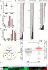
Fig. 1
Fig. 1
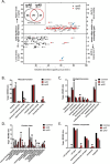
Genomic and proteomic analysis of control, spAD, and rpAD cases. The figure depicts the differences in A age at onset, B proteomic expression, C APOE allele frequency, and D APOE genotype among spAD (grey) and rpAD (red) cases. E spAD and rpAD presented differential expression in 36 and 47 proteins, respectively. F, G The relative expression of these targets in the two variants of AD is also depicted. “*” represents p < 0.05, “***” represents p < 0.001, ns = non-significant
Fig. 2
Fig. 2
Functional characterization of differentially regulated proteins in control, spAD, and rpAD cases. A The relative expression of proteins with significant expression variations and their categorization based on B molecular function, C biological process, D protein class, and E cellular location
Fig. 3
Fig. 3
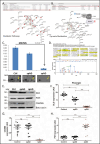
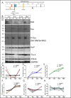
Differential expression of metabolic proteins, especially DLDH, in rpAD cases. Functional characterization and network analysis revealed alterations in A metabolic pathways, including B pyruvate metabolism. C The expression of DLDH, as detected by mass spectrometry and illustrated by the corresponding DLDH spot in the 2DE image (D), was significantly reduced in rpAD. Western blot analysis (E) also depicted expression variations in DLDH (F), CAHM6 (G), and PDH (H). Data is presented as mean ± SEM. “**” represents p < 0.01, “***” represents p < 0.001, ns = non-significant
Fig. 4
Fig. 4
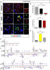
Colocalization of DLDH and Tau. Confocal immunofluorescence imaging (A) depicted the relative expression of DLDH (B) and Tau (C) in brain sections and a significantly higher colocalization between the two in spAD cases (D). Ligplot analysis revealed the residues involved in the interaction of Tau and DLD (E, F). The blue dotted line represents hydrogen bonding, and the red half-circles show DLDH hydrophobic residues. The pink half-circles show Tau hydrophobic residues. Data is presented as mean ± SD. “*” represents p < 0.05, “***” represents p < 0.001, ns = non-significant
Fig. 5
Fig. 5
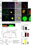
Colocalization of DLDH and Aβ. Confocal immunofluorescence imaging depicted the colocalization of DLDH and Aβ (A, B) in brain sections. A more extensive DLDH internalization in Aβ plaques was observed in rpAD in comparison to spAD brains. Ligplot analysis revealed the residues involved in the interaction of Tau and Aβ (C, D). The blue dotted line represents hydrogen bonding, and the red half-circles show DLDH hydrophobic residues. The pink half-circles show Tau hydrophobic residues
Fig. 6
Fig. 6
In vitro aggregation assays to identify the role of DLDH in Aβ aggregation. Addition of DLDH to the reaction mixture, optimized for the formation of Aβ fibrils, reduced the formation of fibrils as observed by ThT absorbance (A). Similar trends were observed for Aβ25–35 (B), Aβ40 (C), and Aβ42 (D) in negative staining electron microscopy. The plotted data represents two independent experiments
Fig. 7
Fig. 7
Expressional changes in DLDH and other relevant proteins during the progression of AD. 3xTg-AD mice models were developed, and proteins were extracted at 2, 6, 12, and 18 months for immunoblot analysis (A, B). The changes in relative expression of DLDH (C), Tau (D), P-Tau (E), PrP (F), Tia-1 (G), and syntaxin 6 (H) throughout the progression of the disease are depicted. “wt” stands for wild type

References

    1. Alzheimer’s Association, 2023 Alzheimer’s disease facts and figures. Alzheimer’s & Dement 2023;19(4):1598–695 - PubMed
    1. Schmidt C, Redyk K, Meissner B, Krack L, Von Ahsen N, Roeber S, Kretzschmar H, Zerr I (2010) Clinical features of rapidly progressive Alzheimer’s disease. Dement Geriatr Cogn Disord 29(4):371–378 - PubMed
    1. Schmidt C, Haïk S, Satoh K, Rábano A, Martinezmartin P, Roeber S, Brandel JP, Calero-Lara M et al (2012) Rapidly progressive Alzheimer’s disease: a multicenter update. J Alzheimers Dis 30(4):751–756 - PubMed
    1. Hermann P, Haller P, Goebel S, Bunck T, Schmidt C, Wiltfang J, Zerr I (2022) Total and phosphorylated cerebrospinal fluid Tau in the differential diagnosis of sporadic Creutzfeldt-Jakob disease and rapidly progressive Alzheimer’s disease. Viruses 14(2):276 - PMC - PubMed
    1. Younas N, Zafar S, Shafiq M, Noor A, Siegert A, Arora AS, Galkin A, Zafar A et al (2020) SFPQ and Tau: critical factors contributing to rapid progression of Alzheimer’s disease. Acta neuropathol 140:317–339 - PMC - PubMed

LinkOut - more resources