[Preprint]. 2025 Sep 30:rs.3.rs-7330245.
doi: 10.21203/rs.3.rs-7330245/v1.

Integrated Multiclass Driver ctDNA Profiling Enables MPNST Detection and Monitoring in NF1 Patients

Affiliations

Integrated Multiclass Driver ctDNA Profiling Enables MPNST Detection and Monitoring in NF1 Patients

Paul A Jones et al. Res Sq. .

Abstract

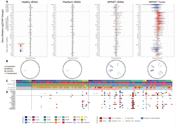
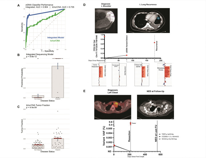
We developed and cross-validated an integrated circulating tumor DNA assay incorporating SNVs, indels, CNAs, and SVs to distinguish neurofibromatosis type 1 patients with malignant peripheral nerve sheath tumors from those with benign plexiform neurofibromas or tumor-free controls. Among 82 participants, the assay achieved an AUC of 0.904, compared with 0.735 for genome-wide CNAs. We also detected recurrent disease-specific driver alterations, relapse up to three months before diagnosis, and early clearance consistent with durable remission.


Conflict of interest statement

Declarations Competing Interests: P.A.J, J.J.S., R.T.S., J.F.S., A.A.C. and A.C.H. have patent filings related to cancer biomarkers. A.A.C. has licensed technology to Droplet Biosciences, Tempus Labs, and LiquidCell Dx. A.A.C. has served as a consultant/advisor to Roche, Tempus, Guardant Health, Exact Sciences, Caris, Geneoscopy, Illumina, Myriad Genetics, Invitae, Daiichi Sankyo, AstraZeneca, AlphaSights, DeciBio and Guidepoint. A.A.C. has received honoraria from Agilent and Illumina, and has received research support from Illumina, Roche and Tempus. A.A.C. has stock options in Geneoscopy, and ownership interests in Droplet Biosciences, LiquidCell Dx and CytoTrace Biosciences. A.C.H. has served on advisory boards for AstraZeneca/Alexion and Springworks Therapeutics. No potential conflicts of interest were disclosed by the other authors.

Figures

Figure 1
Figure 1. Grand Oncoplot reveals key loss of MPNST-associated tumor suppressors and chromosome 8q candidate oncogenes in plasma, recapitulating alterations found in tumor.
A. Copy-number alteration (CNA) landscape. Relative log2 copy number ratio (CNR) across healthy cfDNA, PN cfDNA, MPNST cfDNA, and MPNST tumor. Genes are ordered by relevance: (CDKN2A/CDKN2B, SUZ12, TP53, EED, NF1); chromosome 8q candidate oncogenes (e.g., TCEA1, PLAG1, CHCHD7); and less transformation-associated or clonal hematopoiesis–related genes below. MPNST samples show frequent gains and losses at disease-associated loci; PN and healthy cfDNA show minimal alteration. Blue, loss; red, gain. B. Structural-variant (SV) summary. SVs across cfDNA and tumor samples, including deletions, duplications, inversions, or translocations. MPNST cfDNA shows extensive rearrangements recapitulated in matched tumors; PN and healthy cfDNA show few events. One cfDNA sample with likely chromothripsis was excluded. C. Demographic and clinical annotation block. Clinical and demographic metadata per sample, aligned across panels. “Decade” indicates age at blood draw; “Sex” indicates male or female. “Disease status” differentiates healthy cfDNA, PN cfDNA, MPNST cfDNA, and MPNST tumor. NF1 denotes clinical diagnostic status, with “Indeterminate” for individuals with suggestive but insufficient features. NF1 germline mutation and metastasis status (localized vs. metastatic) are also shown. D. Single-nucleotide variant (SNV) and indel oncoplot. Nonsynonymous SNVs and short indels across cfDNA and tumor samples. Gene order matches the CNA panel. Variants are classified by the Precision Analysis for Cancer Tracking (PACT) integrating SIFT/PolyPhen as truncating, splice site, or missense; missense variants are further categorized as protein-disrupting, variant of uncertain significance (VUS), or non-disruptive. Only high-confidence calls are shown; germline, clonal hematopoiesis of indeterminate potential (CHIP)-associated, and low-confidence variants were excluded. “Multi-hit” labels mark genes with multiple events per sample. MPNST samples harbor frequent alterations in core suppressors; PN and healthy samples show few pathogenic mutations. Abbreviations: cfDNA, cell-free DNA; CHIP, clonal hematopoiesis of indeterminate potential; CNA, copy number alteration; CNR, copy number ratio; indel, insertion/deletion; MPNST, malignant peripheral nerve sheath tumor; PN, plexiform neurofibroma; SNV, single nucleotide variant; SV, structural variant; VUS, variant of uncertain significance.
Figure 2
Figure 2. Integrated multiclass circulating tumor DNA (ctDNA) classifier outperforms tumor fraction and enables longitudinal tracking of molecular clearance and recurrence.
A. ROC curve. Comparison of the integrated model, integrating single nucleotide variant (SNV), copy number alteration (CNA), and structural variant (SV) features, with ichorCNA-derived tumor fraction. The integrated model achieved a leave-one-out cross-validated (LOOCV) AUC of 0.904, outperforming ichorCNA (AUC = 0.735). All models were trained and evaluated on the same cfDNA cohort. B. Integrated model separation. Predicted malignancy probabilities by disease status. Each point is a cell-free DNA (cfDNA) sample. MPNST and non-malignant groups show clear separation (P = 9.8 × 10⁻¹², Wilcoxon rank-sum test). Red lines denote medians; bars show mean ± s.e.m. C. ichorCNA tumor fraction. ichorCNA tumor fraction for the same cfDNA cohort. Group separation remains significant (P = 4.4 × 10⁻⁴, Welch t test) but is less distinct than the integrated model, highlighting the value of multiclass feature integration. Estimates used off-target reads. D. Longitudinal relapse case. ctDNA tracking at surgical resection, immediate postoperative plasma, three-month follow-up, and recurrence biopsy. A recurrent TP53 deletion was present at resection, undetectable immediately postoperative, and re-emerged in plasma at the three-month follow-up and in the recurrence biopsy. CNA profiles shifted between postoperative and relapse, mirroring tumor evolution. Molecular relapse was detectable ≈ 80 days before clinical diagnosis. Red symbols indicate variant detected; empty circles indicate variant not detected. E. Longitudinal clearance case. ctDNA profiling in a patient with localized MPNST. Three tumor-informed variants (TERT, CCDC56, NCOA2) were detected in tumor and pretreatment plasma but were absent from postoperative and subsequent plasma samples, indicating complete molecular clearance. The patient remained disease-free for ≥ 12 months. Red symbols indicate variant detected; empty circles indicate variant not detected. Abbreviations: AUC, area under the curve; CNA, copy number alteration; cfDNA, cell-free DNA; ctDNA, circulating tumor DNA; ichorCNA, tumor fraction estimation from copy number analysis; LOOCV, leave-one-out cross-validation; MPNST, malignant peripheral nerve sheath tumor; PN, plexiform neurofibroma; ROC, receiver operating characteristic; s.e.m., standard error of the mean; SNV, single nucleotide variant; SV, structural variant.

References

    1. Porter D. E., Prasad V., Foster L., Dall G. F., Birch R., & Grimer R. J. (2009). Survival in Malignant Peripheral Nerve Sheath Tumours: A Comparison between Sporadic and Neurofibromatosis Type 1-Associated Tumours. Sarcoma, 2009, 756395. 10.1155/2009/756395 - DOI - PMC - PubMed
    1. Nguyen R., Dombi E., Widemann B. C., Solomon J., Fuensterer C., Kluwe L., Friedman J. M., & Mautner V. F. (2012). Growth dynamics of plexiform neurofibromas: a retrospective cohort study of 201 patients with neurofibromatosis 1. Orphanet journal of rare diseases, 7, 75. 10.1186/1750-1172-7-75 23035791 - DOI - PMC - PubMed
    1. Wasa J., Nishida Y., Tsukushi S., Shido Y., Sugiura H., Nakashima H., & Ishiguro N. (2010). MRI Features in the Differentiation of Malignant Peripheral Nerve Sheath Tumors and Neurofibromas. American Journal of Roentgenology, 194(6), 1568–1574. 10.2214/Ajr.09.2724 - DOI - PubMed
    1. Ahlawat S., Blakeley J. O., Rodriguez F. J., & Fayad L. M. (2019). Imaging biomarkers for malignant peripheral nerve sheath tumors in neurofibromatosis type 1. Neurology, 93(11), e1076–e1084. 10.1212/WNL.0000000000008092 - DOI - PubMed
    1. Carton C., Evans D. G., Blanco I., Friedrich R. E., Ferner R. E., Farschtschi S., Salvador H., Azizi A. A., Mautner V., Röhl C., Peltonen S., Stivaros S., Legius E., Oostenbrink R., & ERN GENTURIS NF1 Tumour Management Guideline Group (2023). ERN GENTURIS tumour surveillance guidelines for individuals with neurofibromatosis type 1. EClinicalMedicine, 56, 101818. 10.1016/j.eclinm.2022.101818 - DOI - PMC - PubMed

Publication types

LinkOut - more resources