A Concept for MRI-Based Cholesteatoma Detection in Cochlear Implant Recipients
- PMID: 41283505
- PMCID: PMC12642017
- DOI: 10.3390/audiolres15060162
A Concept for MRI-Based Cholesteatoma Detection in Cochlear Implant Recipients
Abstract
Introduction: Cochlear implantation is the treatment of choice for severe hearing loss and deafness. Cholesteatomas can cause this deafness. A frequently used procedure in the course of surgical rehabilitation is a subtotal petrosectomy combined with a cochlear implant. The clinical follow-up of residual cholesteatomas is related to the blind sac closure difficult. Cholesteatoma MRI sequence-related CI magnet artefacts make follow-up challenging. Recent developments in combining cochlear implants and necessary MRI examinations enable the assessment of the internal auditory canal and cochlea. The study aimed to develop a procedure for detecting cholesteatomas in patients with cochlear implants using magnetic resonance imaging (MRI).
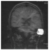
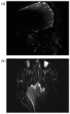
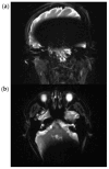
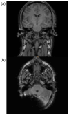
Methods: Ex vivo MRI examinations were performed on five volunteers with fixed cochlear implants (Medel Synchrony) and swim caps. MRI examinations were performed at 1.5 T and 3 T using EPI, HASTE, and RESOLVE sequences (Siemens). The position of the implant was 12 cm distal to the external auditory canal, with anteversional head position of the volunteers in the MRI.
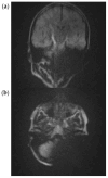
Results: Due to artefact effects, assessment of the ipsilateral and contralateral mastoid is not possible with EPI sequences and a cochlear implant. The combination of cholesteatoma-detecting MARS sequences (HASTE, RESOLVE), a distal implant position, and a specific head position allows the assessment of the ipsilateral mastoid.
Conclusions: Postoperative cholesteatoma assessment after CI implantation and subtotal petrosectomy appears to be possible under 1.5 T and 3 T, considering the MRI sequence, implant position, and head position.
Keywords: CI; MRI; cholesteatoma.
Conflict of interest statement
The authors declare no conflict of interest.
Figures
References
-
- van de Heyning P., Mertens G., Topsakal V., de Brito R., Wimmer W., Caversaccio M.D., Dazert S., Volkenstein S., Zernotti M., Parnes L.S., et al. Two-phase survey on the frequency of use and safety of MRI for hearing implant recipients. Eur. Arch. Oto-Rhino-Laryngol. 2021;278:4225–4233. doi: 10.1007/s00405-020-06525-3. - DOI - PMC - PubMed
LinkOut - more resources
Full Text Sources
Miscellaneous

