. 2025 Aug 8;2(3):ugaf028.
doi: 10.1093/narmme/ugaf028. eCollection 2025 Jul.

The adaptability of the fungal tRNA ligase's ATP-binding pocket: a potential target for new antifungal drugs

Affiliations

The adaptability of the fungal tRNA ligase's ATP-binding pocket: a potential target for new antifungal drugs

Alastair G McEwen et al. NAR Mol Med. .

Abstract

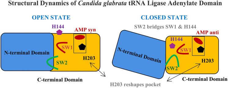
The transfer RNA (tRNA) ligase (TRL1) is a highly conserved multidomain protein that is the archetype of the recently characterized Rnl6 clade. This clade distinguishes itself through a distinct C-terminal domain that sets it apart from other RNA ligase families. TRL1 is an essential component of pre-tRNA splicing and the processing of the Ire1p (Inositol-requiring enzyme 1)-dependent noncanonical splicing of the messenger RNA (mRNA) coding for HAC-1 (Homologous to Activating Transcription Factor / cAMP Response Element-Binding Protein 1 (ATF/CREB1), a transcription factor critical for the unfolded protein response (UPR) in the kingdom of fungi. Here, we report the crystal structure of the N-terminal adenylyl transferase domain (LIG) from Candida glabrata (Nakaseomyces glabratus). The asymmetric unit contained two molecules in complex with noncovalently linked adenosine monophosphate (AMP), revealing conformational differences. In comparison to previous studies, we observe two distinct and partially overlapping ligand-binding pockets, implying new specific residues involved in ligand binding and recognition. These insights on TRL1's ligand adaptability have important implications for the development of targeted therapies.


Conflict of interest statement

None declared.

Figures

Graphical Abstract
Graphical Abstract
Figure 1.
Figure 1.
TRL1 LIG domain primary sequences and secondary structures from CAGLA, CHTHE, and SACER. Secondary structural elements determined in this work and prior investigations [7, 8] are denoted by the letters “S” for β-strands and “H” for α-helices. The black columns represent conserved residues, whereas the gray columns represent similar residues. The nucleotidyltransferase motifs I, Ia, III,IV, and V [10] are underlined. The switch areas are denoted by the symbol “#”. The “A” denotes the necessary LIG SACER residues for ligase function in vivo, as established by alanine scanning mutagenesis (see “Results” section). The asterisks (*) indicate the residues directly interacting with the ATP ligand [10].
Figure 2.
Figure 2.
Structural differences between the two protomers in the crystallographic asymmetric unit. (A) The molecules were aligned and superimposed with matchmaker by matching graphs built on the protein’s secondary-structure elements following an iterative three-dimensional alignment of protein backbone C-α atoms, using as a reference the N-terminal domain [7, 8]. The open and closed state are coloured, respectively, in cyan and yellow; while the CHTHE APK complex (6NOV) is colored in gray. The polygonal figures are explained in the result section. (B) Cartoon representation of the CAGLA LIG domain conformation in its “open” state (molA). Black broken lines represent hydrogen bonds, and salt bridges, and green broken lines represent van der Waals interactions. The residue’s hue varies from cyan (less conserved) to purple (more conserved). Carbon atoms are represented in purple, oxygen atoms in red, nitrogen atoms in blue, and phosphorus atoms in orange. The K110 carbon atoms are depicted in yellow. (C) Cartoon representation of the CAGLA LIG domain in its “closed” state conformation (molB). The K110 carbon atoms are depicted in yellow. (D) Cartoon representation of the CHTHE LIG domain (6N0V). The APK residue, carbon atoms are depicted in yellow. Secondary structural elements are denoted as “h” for α-helices, and “SW” for the switching sites.
Figure 3.
Figure 3.
Conformations of the bound ligands. The ligand AMP in molA (A) and molB (B) are shown in their Polder [17] OMIT (mFo-DFc) map contoured at 3σ. Ligands are represented as sticks, with carbon atoms in yellow or cyan and oxygen atoms in red, nitrogen atoms blue, and phosphorus atoms orange.
Figure 4.
Figure 4.
Structural comparison of the ATP binding pocket of Rnl6 ligases LIG domain. Atomic contacts are represented by dashed lines: hydrogen bonds are colored in black, van der Waals interactions in green. The color of the residue ranges from cyan (less conserved) to purple (more conserved). Carbon atoms are represented in purple, oxygen atoms in red, nitrogen atoms in blue, and phosphorus atoms in orange. Carbon atoms are illustrated in yellow for the lysine residue that becomes covalently bonded to AMP during the ligation. In green are the carbon atoms of the histidine residue described as the “gatekeeper”. (A) The AMP molecule complexed with CAGLA LIG in its “open” state (molA). AMP, shown as sticks, in the intermediatesyn conformation. (B) The AMP molecule complexed with CAGLA LIG in its “closed” state (molB). AMP, shown as sticks, in the anti conformation. (C) The APK intermediate of the CHTHE LIG complex (PDB ID: 6N0V). (D) The Michaelis–Menten AMPcPP complex of the CHTHE LIG complex (PDB ID: 6N67).

References

    1. Pierce CG, Lopez-Ribot JL. Candidiasis drug discovery and development: new approaches targeting virulence for discovering and identifying new drugs. Expert Opin Drug Discovery. 2013; 8:1117–26. 10.1517/17460441.2013.807245. - DOI - PMC - PubMed
    1. Phizicky EM, Consaul SA, Nehrke KWet al.. Yeast tRNA ligase mutants are nonviable and accumulate tRNA splicing intermediates. J Biol Chem. 1992; 267:4577–82. 10.1016/S0021-9258(18)42872-4. - DOI - PubMed
    1. Sidrauski C, Cox JS, Walter P. tRNA ligase is required for regulated mRNA splicing in the unfolded protein response. Cell. 1996; 87:405–13. 10.1016/S0092-8674(00)81361-6. - DOI - PubMed
    1. Phizicky EM, Schwartz RC, Abelson J. Saccharomyces cerevisiae tRNA ligase. Purification of the protein and isolation of the structural gene. J Biol Chem. 1986; 261:2978–86. 10.1016/S0021-9258(17)35882-9. - DOI - PubMed
    1. Apostol BL, Westaway SK, Abelson Jet al.. Deletion analysis of a multifunctional yeast tRNA ligase polypeptide. Identification of essential and dispensable functional domains. J Biol Chem. 1991; 266:7445–55. 10.1016/S0021-9258(20)89467-8. - DOI - PubMed

LinkOut - more resources