. 2025 Nov 28;11(1):552.
doi: 10.1038/s41420-025-02852-8.

Sibiriline, a novel dual inhibitor of necroptosis and ferroptosis, prevents RIPK1 kinase activity and (phospho)lipid peroxidation as a potential therapeutic strategy

Affiliations

Sibiriline, a novel dual inhibitor of necroptosis and ferroptosis, prevents RIPK1 kinase activity and (phospho)lipid peroxidation as a potential therapeutic strategy

Claire Delehouzé et al. Cell Death Discov. .

Abstract

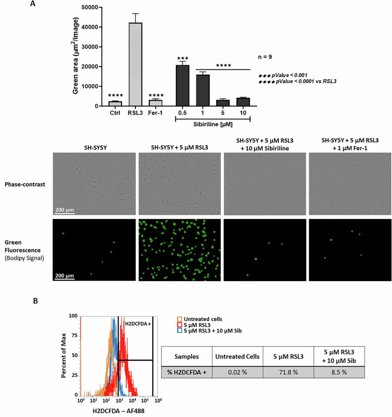
In the past two decades, various non-apoptotic pathways of regulated cell death have been identified; a small subset of these, including necroptosis and ferroptosis, manifests the phenotypic features of necrotic death. These two regulated necroses are being extensively studied because of their putative roles in severe acute and chronic pathologies. Moreover, as these regulated necrotic pathways are coactivated in a number of common pathologies, the development of multi-target directed ligands (that is, the use of a polypharmacological strategy) is a path-breaking avenue of research. In this study, we determined that the 7-azaindole derivative, sibiriline, inhibited both RIPK1-driven necroptosis (induced by Tumor Necrosis Factor-α) and ferroptosis (triggered by various classes of ferroptosis inducers), with EC50s against each in the µM range. We next performed a combined large-scale transcriptomic study in order to determine the molecular mechanisms of action of sibiriline. We identified the stress response protein heme oxygenase-1 (HMOX1) as the main biomarker of ferroptosis inhibition by sibiriline. We hypothesized that this compound reacts as an antioxidant to block ferroptosis; indeed, we found that sibiriline inhibits lipid peroxidation by trapping phospholipid-derived peroxyl radicals as a radical-trapping antioxidant (RTA). Taken together, these results show that sibiriline is a new dual inhibitor of necroptosis and ferroptosis cell death pathways; it works by inhibition of both RIPK1 kinase and (phospho)lipid peroxidation. We also demonstrate the in vitro efficacy of sibiriline to inhibit cell death in cell-based models of Parkinson's disease and cystic fibrosis. These findings shed light on the high therapeutic potency of RIPK1 inhibitors with RTA activity.


Conflict of interest statement

Competing interests: Claire Delehouzé, Marie-Thérèse Dimanche-Boitrel, Morgane Rousselot and Stéphane Bach are the cofounders, shareholders and members of the scientific advisory board of SeaBeLife Biotech, which is developing novel therapies for treating liver acute disorders and retinal diseases. Peter Goekjian is member of the scientific advisory board of SeaBeLife Biotech. The other authors declare that they have no competing interests. Ethics approval and consent to participate: Patient data and tissue collection were performed under the agreement with the European Network of Research Ethics Committees and French ethic law. The ethical committee, according to the Medical Research Involving Human Subjects Act, reviewed and approved the study. Informed voluntary consent was obtained from every participant of the study: agreements CHU 19 244 C and CNRS 205782. Regarding consent, all patients are INFORMED about the use of their cells for research purposes. Patients could withdraw their consent at any time, leading to the prompt disposal of their tissue and any derived material.

Figures

Fig. 1
Fig. 1
Structures of sibiriline (Sib, 1) and previously reported dual necroptosis/ferroptosis inhibitors, necrostatin-1 (Nec-1, 2) [24, 50], necrostatin-1f (Nec-1f, 3) [26], nigratine (6E11, 4) [25], KW-2449 (5) [27] and Dovitinib (12) [28].
Fig. 2
Fig. 2. Sibiriline (1) is an inhibitor of ferroptosis cell death.
A SH-SY5Y cells were treated with either 10 µM Erastin, 5 µM RSL3, 10 µM FIN56 or 25 µM FINO2, and increasing concentrations of Sib up to 50 µM or until reaching maximal cellular viability or 1 µM of ferrostatin-1 (Fer-1/F1) as a control. Cell viability was determined after 24 h of treatment, using the MTS assay. The bar graph represents the mean of two replicates. B SH-SY5Y cells were co-treated for 24 h with 5 µM RSL3 and increasing concentrations of Sib or Fer-1. Cell death was estimated by the lactate dehydrogenase (LDH) release assay. Results are plotted in % of LDH release measured in cells treated with RSL3 alone (left axis, colored blue). Cell viability was evaluated by MTS reduction assay. Results obtained (colored red) were plotted as % of maximal viability with DMSO-treated cells (right axis). Data are shown as the mean +/- SEM of three replicates. C NIH3T3 cells were treated with either 1 µM RSL3, 5 ng/ml TNFα and 20 µM z-VAD.fmk (TZ) or a combination of both treatment (TZ + RSL3) and 10 µM of Sib or Nec-1f, 30 µM Nec-1s or 1 µM of Fer-1. Cell viability was evaluated by MTS reduction assay after 16 h of treatment. Data are shown as the mean ± SEM of three replicates of two independent experiments. D NIH3T3 cells were treated with 5 ng/ml TNFα, 20 µM z-VAD.fmk and 1 µM RSL3 and increasing concentrations of Sib or Nec-1f. Cell viability was determined after 16 h of treatment using an MTS assay. Data are shown as the mean ± SEM of two replicates. EC50 values were calculated using graphpad prism software. Statistical analysis was performed using two-way ANOVA and Tukey’s multiple comparisons test using graphpad prism software. *p < 0.05, **p < 0.01, ***p < 0.001 and ****p < 0.0001 vs controls.
Fig. 3
Fig. 3. Sibiriline (1) reduces hallmarks of ferroptosis cell death.
A Lipid peroxidation was detected by cellular BODIPY 581/591 C11 staining. Fluorescence was recorded with the IncuCyte S3 live cell imaging apparatus. Data are shown as the mean ± SEM of nine replicates. Representative phase-contrast and fluorescence images of SH-SY5Y cells stained with BODIPY 581/591 C11 probe, visualized using the IncuCyte S3 live-cell imaging system. B SH-SY5Y cells treated with DMSO or with 5 µM RSL3 with or without 10 µM Sib were stained with H2DCFDA dye. H2DCFDA fluorescence signal, indicative of intracellular ROS level, was measured in the Alexa Fluor 488 channel (AF488) using an AttuneTM NxT flow cytometer. Representative histograms depicting the ROS production in RSL3-treated SH-SY5Y cells (red), Sib + RSL3-treated cells (blue) or vehicle only (DMSO) cells (orange). The analysis was performed with 105 cells for each condition and the percentage were calculated with AttuneTM NxT software (Thermo Fisher).
Fig. 4
Fig. 4. Sibiriline (1) modulates transcription of ferroptosis-related genes.
A Simplified representation of the workflow used for the transcriptomic-based study. B Overlapping of differentially expressed genes (DEGs) in SH-SY5Y cells treated with 5 µM RSL3 vs DMSO (labeled as RSL3, blue circle) and cells treated with RSL3 + Sib (20 µM) vs RSL3 (labeled as Sib + RSL3, red circle). Heatmap showing the 20 most RSL3-DEG genes in RSL3, Sib+RSL3 (Comb) or DMSO treated cells. C Overlapping of DEGs in RSL3 and Sib+RSL3 treated cells with 60 ferroptosis-related genes. The heatmap shows the mRNA expression profile of the five selected genes in RSL3, Sib+RSL3 (Comb) or DMSO treated cells. The lower panel shows the representative histograms of mRNA expression for each of the five genes in each experimental condition.
Fig. 5
Fig. 5. Sib (1) and Sib-f (6) suppress phospholipid peroxidation via radical-trapping antioxidant (RTA) activity.
A The FENIX assay is based upon the DTUN-initiated co-autoxidation of membrane phospholipids and STY-BODIPY, which is used as a signal carrier to enable reaction monitoring by fluorescence. B Radical-trapping stoichiometry (n) and apparent inhibition rate constant (kinh) can be determined directly from reaction progress data from eqs 1 and 2. C Sibiriline (1) and (D) sibiriline-f (6) inhibited FENIX co-autoxidations. Phosphatidylcholine liposomes (1 mM) in the presence of STY-BODIPY (1 µM) initiated with DTUN (0.2 mM) inhibited with 4-32 µM of Sib (1) or Sib-f (6). E Chemical structures of RTAs used in the FENIX assay. Quintessential phenolic RTA, 2,2,5,7,8-pentamethychroman-1-ol (PMC) (gray), used in the determination of the rate of initiation (Ri), sibiriline (blue) and sibiriline-f (red). F Summary of apparent inhibition rate constants (kinh) and radical-trapping stoichiometry (n) derived using eqs 1 and 2. G Evaluation of lipid peroxidation-inhibiting activity of sibiriline (1) and its derivatives by FENIX assay. The RTA effect is expressed in kinh liposomes (see [32] for details). Values reported in the table were determined from the curves reported in Fig S4. n.e.: no significant effect detected up to 32 µM.
Fig. 6
Fig. 6. Efficacy of the identified compounds against Cumen Hydroperoxide-induced cell death in healthy or Cystic Fibrosis (CF) nasal epithelial cells.
AC. LDH release in primary Human Nasal Epithelial Cells (HNECs) from healthy or cystic fibrosis patients exposed to 50 µM of Cumene Hydroperoxide (CuOOH, 50 µM, 8 h) in presence/absence of various concentration of inhibitory compounds. Here Fer1 stands for ferrostatin-1 and was used as internal inhibitory control of CuOOH-induced cell death. The molecules sibiriline (Sib) (1), sibiriline-f (Sib-f) (6) and AC1610F1 (11) are described along the manuscript. ****P ≤ 0.0001, 2-way Anova with multiple comparisons. Values are expressed as mean ± SEM from one experiment (in triplicate) from one independent donor (d1, CF) performed at least three times.
Fig. 7
Fig. 7. Sib (1) protects neurons in a Parkinson model of 6-OHDA-induced rat dopaminergic neurons degeneration.
Primary rat dopaminergic neurons were exposed to 20 µM of 6-OHDA and several doses of Fer-1 or Nec-1s (A) or Sib (C) or a combination of Fer-1 + Nec-1s (B) for 48 h. Cells were fixed and labeled with antibody against tyrosine hydroxylase (TH). Results were expressed as a percentage of the number of TH positive dopaminergic neurons compared to the control non-treated condition. Data were expressed as mean ± SEM of six replicates. A global analysis of the data was performed using one-way ANOVA following by Dunnett’s test. The level of significance is set at p < 0.05.

References

    1. Galluzzi L, Vitale I, Aaronson SA, Abrams JM, Adam D, Agostinis P, et al. Molecular mechanisms of cell death: recommendations of the Nomenclature Committee on Cell Death 2018. Cell Death Differ. 2018;25:486–541. - PMC - PubMed
    1. Degterev A, Hitomi J, Germscheid M, Ch’en IL, Korkina O, Teng X, et al. Identification of RIP1 kinase as a specific cellular target of necrostatins. Nat Chem Biol. 2008;4:313–21. - PMC - PubMed
    1. Delanghe T, Dondelinger Y, Bertrand MJM. RIPK1 Kinase-dependent death: a symphony of phosphorylation events. Trends cell Biol. 2020;30:189–200. - PubMed
    1. Mifflin L, Ofengeim D, Yuan J. Receptor-interacting protein kinase 1 (RIPK1) as a therapeutic target. Nat Rev Drug Discov. 2020;19:553–71. - PMC - PubMed
    1. Degterev A, Ofengeim D, Yuan J. Targeting RIPK1 for the treatment of human diseases. Proc Natl Acad Sci USA. 2019;116:9714–22. - PMC - PubMed

LinkOut - more resources