[Preprint]. 2025 Nov 17:rs.3.rs-7886236.
doi: 10.21203/rs.3.rs-7886236/v1.

Molecular basis for CSB stimulation of the SNM1A DNA repair nuclease

Molecular basis for CSB stimulation of the SNM1A DNA repair nuclease

Peter McHugh et al. Res Sq. .

Abstract

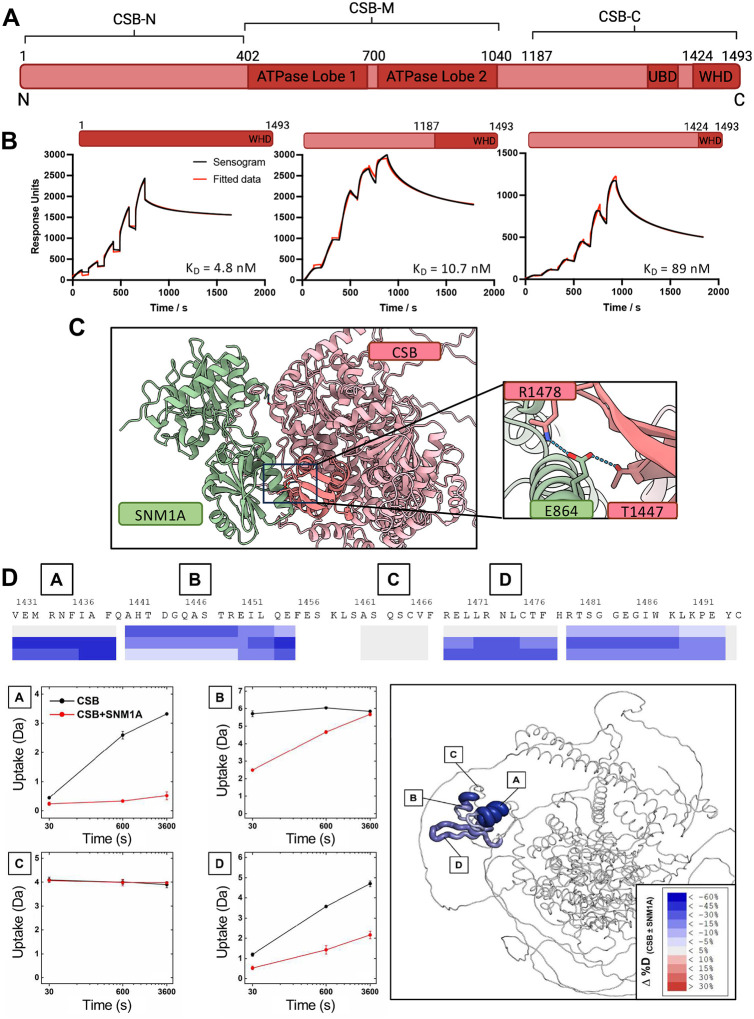
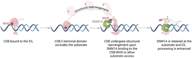
The Cockayne Syndrome B (CSB, ERCC6) protein, interacts with the exonuclease SNM1A during transcription-coupled DNA interstrand (ICL) repair, with CSB facilitating localisation of SNM1A to ICL damage. The functional and mechanistic details of this interaction in DNA repair, however, have not been defined. Here, we demonstrate that CSB enhances SNM1A resection through ICLs and identify a specific interaction between the winged-helix domain of CSB and the nuclease core of SNM1A that is crucial for recruitment and enhancement of nuclease degradation. Biochemical and single-molecule studies on DNA containing sitespecific ICLs reveal that CSB increases the affinity of SNM1A to damaged DNA substrates and also alters the substrate conformation to enhance ICL processing by SNM1A. Notably, CSB was observed preferentially as a dimer when colocalised with SNM1A at ICLs, constrasting with its monomeric nature observed during repair initiation in classical transcription-coupled nucleotide excision repair. The combined results provide molecular insights into the basis of a direct contribution of CSB to a DNA repair reaction.


Figures

Figure 1:
Figure 1:. Defining the molecular interaction between CSB and SNM1A.
A. Domain map of CSB, defining the N-terminal (CSB-N), ATPase (CSB-M) and C-terminal (CSB-C) domains. B. Single-cycle response curves from SPR analyses, with apparent equilibrium dissociation constants given. Parameters determined from fitting are given in Table S1. C. AlphaFold3 modelling of CSB (full-length, a.a. 1–1493, red) and SNM1A (C-terminal domain, a.a. 698–1040, green) with hydrogen bonding interaction between CSB-WHD and SNM1A highlighted. D. HDX-MS of CSB interaction with SNM1A. Differential deuterium uptake was observed in the WHD domain of CSB in the presence SNM1A, exemplified by peptides A, B, and D. No differences between apo- and holo-states were observed for the majority of the protein, an example of which is represented by peptide C. HDX-MS changes were mapped to the AlphaFold model of full-length CSB.
Figure 2:
Figure 2:. Identification of a CSB:SNM1A separation-of function variant.
A. Apparent equilibrium dissociation constants obtained between different CSB (vary a.a. lengths, wild-type and T1447A) and SNM1A (a.a. 698–1040) constructs. The dark red/blue regions of the protein sequence maps represent the amino acid ranges used for experiments. ND = Not determined. B. Multi-cycle SPR analyses of immobilized N-His-ZB-CSB-Avi-Biotin (WT) with SNM1A (WT, a.a. 698–1040, untagged) as the analyte. C. Multi-cycle SPR analyses of immobilized N-His-ZB-CSB-Avi-Biotin (T1447A) with SNM1A (WT, a.a. 698–1040, untagged) as the analyte. D. Co-immunoprecipitation experiment with immobilised GFP or GFP-tagged SNM1A (full-length construct: WT, nuclease-dead variant D736A or E864A). E. Co-immunoprecipitation experiment with immobilised GFP or GFP-tagged CSB (full-length construct: WT or T1447A).
Figure 3:
Figure 3:. CSB enhances SNM1A digestion through ICLs via SNM1A recruitment and substrate modulation.
Oligonucleotides used (with preparation method) are given in Table S3 and Fig. S8. The location of the 3′ 32P is given by a yellow star, Bio represents the location of 5’ biotins on the oligo and 5’P the location of a 5’ phosphate group. Quantification was based upon assays conducted in triplicate; error bars represent one standard deviation. A. Time course assays of SNM1A activity on the ‘0-nucleotide’ ICL substrate in the absence/presence of CSB (WT or T1447A). B. Quantification of the assay shown in B. C. Time course assays of SNM1A activity on single-stranded DNA substrate in the absence/presence of CSB (WT or T1447A). D. Quantification of the assay shown in panel D. E. Time course assays of SNM1A (E864A) activity on single-stranded DNA substrate in the absence/presence of CSB (WT or T1447A). F. Quantification of the assay shown in panel F. data for WT-SNM1A is also shown, from data given in panels B and C, as the assays were run in tandem. G. EMSA of full-length CSB, CSB ATPase domain (CSB-M, a.a. 402–1014), CSB C-terminal domain (CSB-C) a.a., 1187–1493) binding to single stranded DNA in the presence of 4 mM EDTA. H. EMSA of CSB C-terminal domain (a.a. 1187–1493) binding to different DNA structures in the presence of 4 mM EDTA.
Figure 4:
Figure 4:. Investigation of the oligomerisation state of CSB.
A. Mass histograms from mass photometry measurements of the CSB protein. The expected molecular weight of the CSB monomer is 180 kDa. B. Mass histogram of experiments of CSB (expected: 180 kDa, observed: 184 kDa) and SNM1A (expected: 36 kDa, observed: 58 kDa) mixed at 200 nM each, indicating that a 1:1 complex of the two proteins forms. Analysis of the mass counts corresponding to each protein state determined a KD of 499 ± 215 nM (mean ± standard deviation) for the interaction between CSB and SNM1A. C. Fractions from size-exclusion chromatography from co-purification of CSB-C and N-Avi-Biotin-SNM1A (a.a. 698–1040) analysed by SDS-PAGE. D. Size-exclusion chromatography trace from co-purification of CSB-C (a.a. 1187–1493) and SNM1A (a.a. 698–1040). The peak at 12.5–13.5 mL corresponds to a CSB:SNM1A 2:1 complex as supported by SDS-PAGE, MS analysis (for protein identities) and size-exclusion calibration curve from Superdex S200 GL 10/300. See Supplementary Information for calibration curve. E. Size-exclusion chromatography trace from co-purification of FL-CSB and SNM1A (a.a. 698–1040). The peak at 10.5 mL corresponds to the CSB:SNM1A 2:1 complex as supported by SDS-PAGE and MS analysis (for protein identification) and quantitative size-exclusion chromatography (Superdex S200 GL 10/300 column). F. Fractions from size-exclusion chromatography from co-purification of full-length CSB and SNM1A (a.a. 698–1040) analysed by SDS-PAGE. G. Representative kymograph of CSB (2 colour experiment) binding to lambda DNA. H. CRTD analysis of CSB binding to lambda DNA. I. Schematic of the possible modes of DNA binding for two colour protein experiment with two labelled CSB proteins. J. Bar graph showing the representative of different types of molecular assembly events observed with the two colour CSB experiment.
Figure 5:
Figure 5:. Single-molecule studies of CSB:SNM1A assembly on undamaged DNA.
A. Graphical representation of the potential modes of molecular assembly of CSB and SNM1A binding and dissociating, alone or together on DNA. B. Representative kymograph of CSB (JF-635, red) and SNM1A (JF-552, green) binding to lambda DNA. C. Bar graph showing the percentage of events represented by different categories of colocalisation for CSB and SNM1A binding alone or together to lambda DNA. The schematic in panel B is to be used as the key. D. CRTD plot of SNM1A dwell times alone (green) or colocalised with CSB in category 9 events (grey) on lambda DNA. Two-phase binding regimes were determined for both types of events, with average binding lifetimes shown here (individual lifetimes of each regime are given in Table S5). E. CRTD plot of SNM1A dwell times on monomer (grey) or dimer CSB (pink) on lambda DNA i.e., separating the monomer and dimer CSB in category 9 colocalisation events with SNM1A from panel C. Two-phase binding regimes were fit for each with the average of the two lifetimes given. The lifetime of each binding regime is given in Table S5.
Figure 6:
Figure 6:. Single-molecule studies of CSB:SNM1A assembly on ICL-containing DNA.
A. Diagram showing the design of an transcription-coupled ICL repair DNA repair intermediate oligonucleotide that was ligated into two, terminally biotinylated 6.3 kb DNA handles via complementary overhangs. B. Representative kymograph of CSB (JF-635, red) and SNM1A (JF-552, green) binding to the ICL flap substrate. C. CRTD plot of SNM1A dwell times alone (green) or colocalised with CSB in category 9 events (grey) on the ICL. Two-phase binding regimes were determined for colocalised event (with the average given) with both lifetimes given in Table S6. A one-phase binding regime was fit to the SNM1A binding events with the average binding lifetimes shown here. D. Bar graph showing the percentage of events represented by different categories of colocalisation for CSB and SNM1A binding alone or together to the centralized ICL. E. CGTD plot of recruitment times of SNM1A to CSB-bound DNA on dsDNA (black) or the ICL (blue). A two-phase decay was fit to the recruitment of SNM1A to CSB, with the average given here (both given in Table S6) and a single regime fit to SNM1A recruitment to CSB on ICLs. F. Bar graph showing the oligomerization state of all CSB events observed on lambda (ds) DNA or localised at the ICL. Monomer/dimer = CSB event was solely monomeric/dimeric for the entire DNA binding event. Exchange = the CSB oligomerisation state exchanges during DNA binding. G. Bar graph showing the oligomerisation state of CSB when co-localised with SNM1A on lambda (ds) DNA or localised at the ICL.
Figure 7:
Figure 7:
Proposed mechanism of how CSB enhances the ICL processing activity of SNM1A.

References

    1. Hashimoto S., Anai H. & Hanada K. Mechanisms of interstrand DNA crosslink repair and human disorders. Genes and Environment 38, 1–8 (2016). - PMC - PubMed
    1. Rycenga H. B. & Long D. T. The evolving role of DNA inter-strand crosslinks in chemotherapy. Current Opinion in Pharmacology vol. 41 20–26 Preprint at 10.1016/j.coph.2018.04.004 (2018). - DOI - PMC - PubMed
    1. Clauson C., Schärer O. D. & Niedernhofer L. Advances in understanding the complex mechanisms of DNA inter strand cross-link repair. Cold Spring Harb Perspect Med 3, 1–25 (2013).
    1. Housh K. et al. Formation and Repair of an Interstrand DNA Cross-Link Arising from a Common Endogenous Lesion. J Am Chem Soc 143, 15344–15357 (2021). - PMC - PubMed
    1. McHugh P. J. XPF–ERCC1: Linchpin of DNA crosslink repair. PLoS Genet 16, 1–5 (2020).

Publication types

LinkOut - more resources