

Immunofluorescent examination of biopsies from long-term renal allografts
- PMID: 4189456
- PMCID: PMC2765872
- DOI: 10.1056/NEJM197002192820802
Immunofluorescent examination of biopsies from long-term renal allografts
Abstract
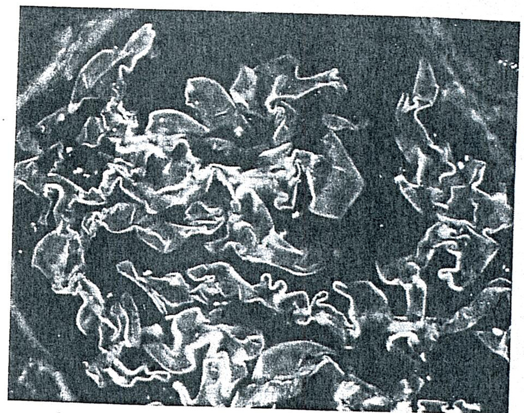
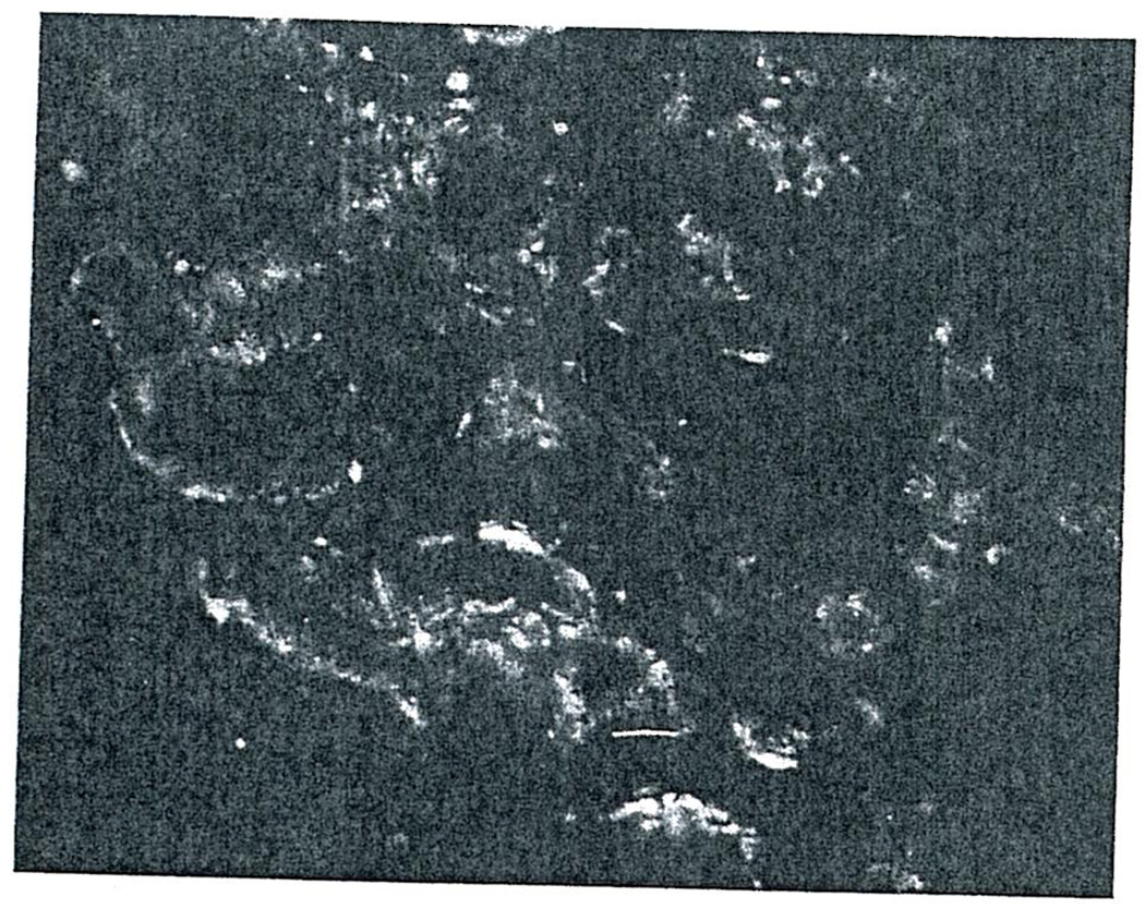
Immunofluorescent examination of open renal biopsies revealed clear-cut glomerular localization of immunoglobulins not related clearly to the quality of donor-recipient histocompatibility in 19 of 34 renal allografts. The biopsies were obtained 18 to 31 months after transplantations primarily from related donors with a variable quality of histocompatibility match. IgG was the predominant immunoglobulin class fixed in 13 biopsies, and IgM in six. The pattern of immunoglobulin deposition was linear, connoting anti-GBM antibody in four of the 19; it was granular and discontinuous, connoting antigen-antibody-complex deposits, in 13. An immune process may affect glomeruli of renal allografts by mechanisms comparable to those that cause glomerulonephritis in native kidneys. The transplant glomerulonephritis may represent a persistence of the same disease that originally destroyed the host kidneys or the consequence of a new humoral antibody response to allograft antigens.
Figures

References
-
- Peterson EW, McPhaul JJ, Jr, McIntosh DA. Serial histologic alterations in human renal homotransplants. Amer J Clin Path. 1966;45:521–532. - PubMed
-
- Fish AJ, Herdman RC, Kelly WD, et al. Glomerular changes in well-functioning human renal homografts. Transplantation. 1967;5:1338–1343. - PubMed
-
- Porter KA, Dossetor JB, Marchioro TL, et al. Human renal transplants. I. Glomerular changes. Lab Invest. 1967;16:153–181. - PubMed
-
- Glassock RJ, Feldman D, Reynolds ES, et al. Human renal isografts: a clinical and pathologic analysis. Medicine (Balt) 1968;47:411–454. - PubMed
MeSH terms
Substances
Grants and funding
LinkOut - more resources
Full Text Sources