. 1973 Oct 9;12(21):4171-9.
doi: 10.1021/bi00745a021.

Quenching of protein fluorescence by oxygen. Detection of structural fluctuations in proteins on the nanosecond time scale

Quenching of protein fluorescence by oxygen. Detection of structural fluctuations in proteins on the nanosecond time scale

J R Lakowicz et al. Biochemistry. .

Abstract

Quenching of the tryptophan fluorescence of native proteins was studied using oxygen concentrations up to 0.13 m, corresponding to equilibration with oxygen at a pressure of 1500 psi. Measurement of absorption spectra and enzymic activities of protein solutions under these conditions reveal no significant perturbation of the protein structure. The oxygen quenching constant (k+*) for a variety of proteins indicates that the apparent oxygen diffusion rate through the protein matrix is 20–50% of its diffusion rate in water. No tryptophan residues appear to be excluded from quenching, and no correlation of the fluorescence emission maxima with k+* was found, indicating that the rapid oxygen diffusion is present in all regions of the protein, even those normally considered inaccessible to solvent. Energy transfer among tryptophans was excluded as a possible mechanism for the rapid quenching by studies using 305-nm excitation, where energy transfer is known to fail. The dynamic character of the observed quenching was proven by the proportional decrease of the fluorescence lifetimes and yields measured under the same conditions. We conclude that proteins, in general, undergo rapid structural fluctuations on the nanosecond time scale which permit diffusion of oxygen.


Figures

FIGURE 1:
FIGURE 1:
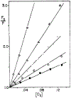
Typical Stern–Volmer plots for proteins. Instrumental conditions used are listed in footnote a of Table II: tryptophan (◯); bovine serum albumin (Δ); IgG (·); aldolase (◻); and α-chymotrypsin(∎).
FIGURE 2:
FIGURE 2:
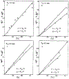
Technical fluorescence emission spectra of proteins equilibrated with air and increased oxygen pressure. The fraction of the original fluorescence remaining is listed as F/F0. Experimental conditions are listed in the footnotes to Table II: (a) human serum albumin; (b) bovine serum albumin; (c) aldolase; (d) carbonic anhydrase.
FIGURE 3:
FIGURE 3:
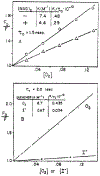
Oxygen quenching of protein fluorescence as observed by fluorescence lifetimes (◯) and yields (Δ). Experimental conditions are listed in the footnotes to Table II and under Experimental Procedures: (a) aldolase; (b) α-chymotrypsin; (c) pepsin; (d) carbonic anhydrase.
FIGURE 4:
FIGURE 4:
(a) Oxygen quenching of lysozyme in the presence (Δ) and absence (◯) of (N-Ac-GlcN)2; 0.1 m sodium phosphate, pH 7; 280-nm excitation, 340- (no (N-Ac-GlcN)2) or 330-nm (with (N-Ac-GlcN)2 emission; [lysozyme] = 7.6 × 10⁻⁵ m, [(N-Ac-GlcN)2] = 3.2 × 10⁻³ m. (b) Comparison of oxygen and iodide quenching of trypsinogen; O2 quench: 0.001 m HC1, 280-nm excitation; I quench: 0.001 m HC1, 290-nm excitation, [KCl] + [KI] = 0.50 m, ca. 10−4 m Na2S2O3; 334-nm emission was used for both samples.
FIGURE 5:
FIGURE 5:
Modified Stern–Volmer plots to determine the fraction of accessible tryptophans in pepsin by iodide (a) or oxygen (b) quenching. Both samples were excited at 290 nm, and the emission intensity recorded at 342 nm: I quench, 0.01 m HCl, [KCl] + [KI] = 0.5 m, ca. 10−4 m Na2S2O3; O2 quench, 0.01 m HC1.

References

    1. Arnon R, and Perlmann GE (1963), J. Biol. Chem 238, 653.
    1. Armstrong McD. J., Myers DV, Verpoorte JA, and Edsall JT (1966),J. Biol. Chem 241, 5137. - PubMed
    1. Bakhshiev NG (1964), Opt. Spectrosc. (USSR) 16,446.
    1. Brand L, and Gohlke JR (1971), J. Biol. Chem 246,2317. - PubMed
    1. Brandts JF, Oliveira RJ, and Westort C (1970), Biochemistry 9,1038. - PubMed

MeSH terms

LinkOut - more resources