. 1973 Aug;137(2):179-99.

The origin, hormonal nature, and action of hepatotrophic substances in portal venous blood

The origin, hormonal nature, and action of hepatotrophic substances in portal venous blood

T E Starzl et al. Surg Gynecol Obstet. 1973 Aug.
No abstract available


Figures

FIG 1
FIG 1
Partial portacaval transposition, a and b, Entire vena caval flow is directed into either the left or the right portal venous branch, c and d, This differs from a and b in that the vena caval flow excludes the adrenal and renal blood. A venous graft is always required to bridge the gap.
FIG 2
FIG 2
Technique of division of splanchnic venous flow into a pancreatico-gastroduodenal-splenic compartment and an intestinal compartment. Blood from these respective sources is directed into the right or left lobes. The tail of the inferior lobe of the pancreas was resected since it drains separately into the mesenteric vein.
FIG 3
FIG 3
Techniques of transposition for the dogs in group 3. a, Standard portacaval transposition of Child, b, Modified transposition which eliminates adrenal and renal venous blood from the portal blood samples. A venous graft is always required.
FIG 4
FIG 4
Hepatocyte shadows traced during histopathologic examination. These were later cut out on standard paper and weighed as an index of hepatocyte size. The specimens depicted were from the experimental group 2 (see Fig. 2a). The right lobes with the large hepatic cells received venous blood from the pancreas, stomach, duodenum, and spleen. The relatively shrunken left lobes with the small hepatocytes received intestinal blood.
FIG 5
FIG 5
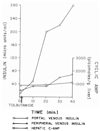
Changes in peripheral and portal venous insulin and. hepatic cyclic 3′, 5′-adenosine monophosphate, cyclic AMP, occurring in a normal dog infused with tolbutamide. Note that the peak insulin response in the portal blood occurred 25 to 40 minutes after infusion and that no significant alterations in hepatic cyclic 3′, 5′-adenosine monophosphate were caused acutely by the tolbutamide itself.
FIG 6
FIG 6
Results with the tolbutamide-glucagon test in three dogs from groups la or 1 c in which there was clotting of the venous grafts. Consequently, the left branch of the portal vein transmitted no blood at all while the right lobes had a full splanchnic inflow. Note that the hepatic cyclic 3′, 5′-adenosine monophosphate response to exogenous glucagon was considerably less in the lobes having a portal inflow compared with the lobes suffering from portal devascularization. The relatively lower cyclic 3′, 5′-adenosine monophosphate presumably represented a restraining insulin effect.
FIG 7
FIG 7
The morphologic consequences, after 2 ± 0.5 (S. D.) months in the dogs of group 1, of supplying parts of the liver with nonhepatic splanchnic venous blood, shaded areas, as compared with vena caval blood. Results are also given for six normal dogs. Note the gross weight gain and hepatocyte hypertrophy on the side perfused with splanchnic blood. Although the relative sizes of the lobes were altered according to the source of portal venous inflow, note that the ratio of the liver weight to total body weight was relatively unchanged. One standard deviation is depicted in the bar graphs and written out for the weight percentages.
FIG 8
FIG 8
The morphologic consequences of splanchnic venous flow division in the dogs in group 2 compared with normal dogs after 28 to 173 days, average 73. The liver fractions which were perfused with venous blood from the pancreatic, gastroduodenal, and splenic areas are shaded. Note that these portions gained weight and underwent an increase in hepatocyte size relative to the other side while the total liver weight to body weight ratios were little altered. One standard deviation is depicted graphically on the bar graphs and written out for the weight percentages.
FIG 9
FIG 9
Photomicrographs of sections from a liver which had been subjected to splanchnic flow division. The part of the liver supplied by the pancreatico-gastroduodenal-splenic blood, panels on left, shows enlargement of the liver lobules, abundant glycogen, and hypertrophy and hyperplasia of hepatocytes when compared with the part supplied by intestinal blood, panels on right. Upper, Reticulin stain, ×10. Middle, Periodic acid-Schiff, ×87. Lower, Hematoxylin and eosin, × 87.
FIG 10
FIG 10
Aminophylline infusion tests in five dogs with partial portacaval transposition. In all but one, the rate of formation of cyclic 3′, 5′-adenosine monophosphate was greater in the lobes deriving their portal inflow from the inferior vena cava although these differences were usually not obvious until several minutes had gone by.
FIG 11
FIG 11
Rate of cyclic 3′, 5′-adenosine monophosphate formation, as measured by the aminophylline infusion test, in the single successful experiment of group 1 d.
FIG 12
FIG 12
Aminophylline infusion tests in three dogs taken from groups la or 1c in which the left portal branch clotted, leaving these lobes supplied only with arterial blood, whereas the right lobes received both arterial and splanchnic venous blood. Note that the rate of synthesis of hepatic cyclic 3′, 5′-adenosine monophosphate was greater in the right lobes having a portal inflow than in the left lobes suffering from total portal devascularization. The time units are in minutes.
FIG 13
FIG 13
Results of tolbutamide-glucagon tests in eight dogs with partial portacaval transposition, demonstrating the effect of endogenous insulin in the lobes receiving splanchnic venous blood. These insulin-controlled lobes had a restrained cyclic 3′, 5′-adenosine monophosphate response to the exogenous glucagon whereas the response in the other lobes was uninhibited.
FIG 14
FIG 14
Aminophylline infusion tests two months after splanchnic flow division in four dogs. Cyclic 3′, 5′-adenosine monophosphate synthesis was much more rapid in the hepatic lobes receiving pancreatic-gastro-duodenal-splenic blood than in the contralateral lobes supplied with intestinal venous effluent.
FIG 15
FIG 15
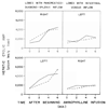
Tolbutamide-glucagon infusion tests in five dogs two months after splanchnic flow division. In the two experiments in which pancreatic-gastroduodenal splenic blood passed to the left lobes, group 2 b, there were no significant differences in the hepatic cyclic 3′, 5′-adenosine monophosphate concentrations on the two sides of the liver. However, in the three dogs of group 2a, top, there was a runaway response in the lobes receiving intestinal venous blood compared with a restrained response in the lobes nourished by pancreatic-gastroduodenal-splenic venous blood.
FIG 16
FIG 16
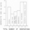
Summary from the different experiments depicted in Figure 1 and Figure 2 shows the influence of the type of portal inflow upon hepatocyte size. The data are taken from Figure 7 and Figure 8 and are compared with values obtained from the livers of normal dogs. Note that the presence of hypertrophy or atrophy was almost exclusively under the control of pancreatic-gastroduodenal-splenic blood and that the addition or subtraction of blood from other sources was of little further consequence.

References

    1. Albano O, Francavilla A. Effect of CC14 intoxication in the synthesis of β-Iipoprotein fractions in rat liver. Clin. Chim. Acta. 1968;19:363. - PubMed
    1. Barron EJ, Hanahan DJ. Observations on the silicic acid chromatography of the neutral Iipides of rat liver, beef liver, and yeast. J. Biol. Chem. 1958;231:493. - PubMed
    1. Bergen SS, Hilton JG, Van Itallie TB. Glycogenolytic effect of adenosine 3′–5′-monophosphate in the canine liver. Endocrinology. 1966;79:1065. - PubMed
    1. Bloom WL, Lewis GT, Schumpert MZ, Shen T-M. Glycogen fractions of liver and muscle. J. Biol. Chem. 1951;188:631. - PubMed
    1. Bocher NLR. Experimental aspects of hepatic regeneration. N. Engl. J. Med. 1967;277:686. - PubMed

MeSH terms

LinkOut - more resources