. 1974 Mar;7(2):229-49.
doi: 10.1016/0026-2862(74)90008-9.

Microvasculature of the dog left ventricular myocardium

Microvasculature of the dog left ventricular myocardium

J B Bassingthwaighte et al. Microvasc Res. 1974 Mar.

Abstract

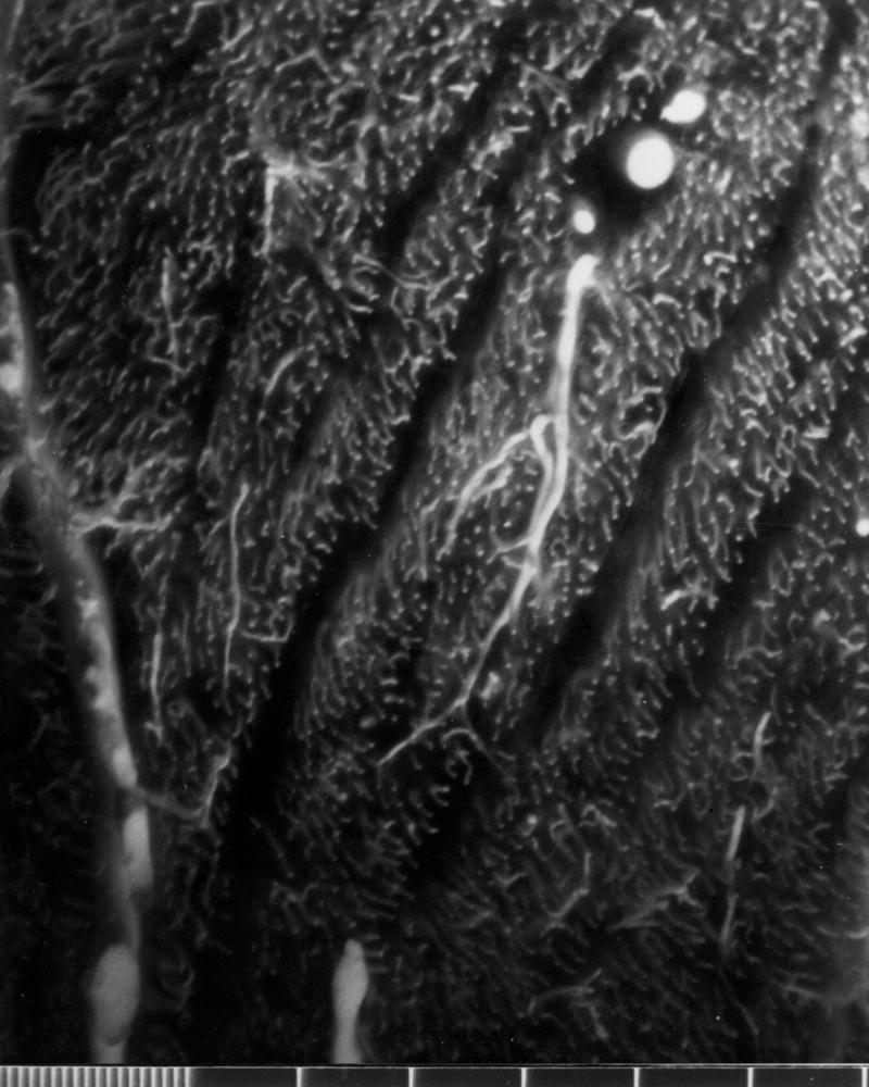
One of the main branches of the left main coronary artery of normally beating dog hearts was perfused with a silicone elastomer which solidified within the vasculature. Prolonged immersion in increasingly concentrated ethanol and in methyl salicylate rendered the tissue translucent and the vasculature clearly visible. Surfaces were photographed by reflected or transmitted light microscopy, showing large groups of capillaries running parallel to muscle fibers and extending for up to a few centimeters. The arrangement of arteriolar inflows to the capillary network and venular outflows (two to four times as frequent) suggested that functional capillary lengths were 500–1000 μm. Estimates of capillary diameters, presumably at maximal dilatation, were 5.6 ± 1.3 μm. Capillary densities within muscle groups were 3100–3800/mm2, giving intercapillary distances of 19–17.5 μm. With the lesser density value, the capillary surface area is estimated to be 500 cm2/g of myocardium. Inclusion of interfascial spaces lowered the average density to about 2500/mm2. Unbranched capillary lengths averaged 100 μm, with a strongly right-skewed distribution. The anatomic arrangement provides a basis mainly for concurrent flow in neighboring capillaries, and also for some diffusional exchange between inflow and outflow regions.


Figures

Fig. 1
Fig. 1
Shrinkage on immersion plotted as a function of sequentially higher concentrations of ethanol and methylsalicylate. A: Shrinkage of myocardium in the form of 1–2 ml pieces (closed symbols). Ordinate is M/M0, measured mass divided by initial mass of the same heart or piece, or V/V0, volume over initial volume. Bars give+SD. (Densities of 100% alcohol and methylsalicylate are 0.79 and 1.17 g/ml. The density of the whole hearts decreased from an initial average of 1.06 g/ml to 0.88 in 100% ethanol and in methylsalicylate increased to 1.18 g/ml.) B. Diameter of columns of Microfil within capillaries.
Fig. 2
Fig. 2
Vasculature of the left ventricular epicardium filled with Microfil. Scale divisions are 1 mm. The top is toward the base of the heart. The capillaries form dense parallel networks in the middle upper region running 30° to the right of the ventricular axis and in the lower half running 30° to the left. Most of the larger vessels are veins. The arrow tips mark arterioles.
Fig. 3
Fig. 3
Venules and capillaries on left ventricular epicardial surface. Scale divisions are 10 and 100 μm. (Different heart from that shown in Fig. 2.) No arterioles are present.
Fig. 4
Fig. 4
Subepicardial vasculature. Section parallel to epicardium, 1 mm deep. Scale divisions 10 and 100 μm. A 60-μm arteriole, accompanied by two venules, gives rise to three 10-μm arterioles, two short and one long (at arrow). The insert (same scale) shows a 160-μm vein giving rise to small venules and branching rapidly into the parallel capillaries.
Fig. 5
Fig. 5
Sections of ventricular myocardium near apex stained with hematoxylin and eosin (heart C). Scale shows 10-μm divisions. Sarcomere lengths are distinctly shortened. Note the separations between individual fibers and groups of fibers.
Fig. 6
Fig. 6
Frequency histogram of uncorrected capillary diameters measured on histologic sections by transmission microscopy. The ordinate is the percentage of observations per 1-μm class width so that the area under each curve is unity. Class width for plotting = 0.2 μm. (Numbers of observations are given in parentheses.) Measurements from cross sections, dashed line, where minor diameters are taken from any elliptical sections (e.g., left panel, Fig. 5), give lower estimates than those from longitudinal sections (e.g., right panel of Fig. 5), dotted line, where one tends to avoid measuring at points which might be too thin because of sectioning partly through the capillary itself. All data = large dots.
Fig. 7
Fig. 7
Cross section of endocardial region of left ventricle. At right are a 15m arteriole (and a branch cut off from it) with two other vessels, presumably venules. Scale divisions are 10 and 100 μm. Overall capillary density here was about 3000 ± 600/mm2 in 90-μm squares, but was higher (3300 + 530/mm2) in smaller wholly muscular regions.
Fig. 8
Fig. 8
Apical left ventricular myocardium in longitudinal section viewed from the endocardial side. An arteriole and branching venule are visible within the tissue lying toward the epicardial surface.
Fig. 9
Fig. 9
Two venules and visceral pericardium of the left ventricular surface. Scales are 10 and 100 μm. An arteriole lies deep. The diagonal branch at the lower right is an intervenous connection joining these two venules. The figure is a composite of two photographs of adjacent regions. (There is an artefact in the left lower corner that should be ignored.)

References

    1. Bassingthwaighte JB. A concurrent flow model for extraction during transcapillary passage. Circ Res. 1974 accepted. - PMC - PubMed
    1. Bassingthwaighte JB, Strandell T, Yipintsoi T. Flow limited washout of diffusible solutes from the heart. In: Crone C, Lassen NA, editors. Capillary Permeability. Munksgaard; Copenhagen: 1970. pp. 580–585.
    1. Bassingthwaighte JB, Yipintsoi T. Countercurrent exchange of water in the heart. Biophys J. 1969;9:232A.
    1. Detar R, Bohr DF. Oxygen and vascular smooth muscle contraction. Amer J Physiol. 1968;214:241–244. - PubMed
    1. Fabel H. Normal and critical O2 supply of the heart. In: Lubbers DW, Luft UC, Thews G, Witzleb E, editors. Oxygen Transport in Blood and Tissue. Georg Thieme Verlag; Stuttgart, Germany: 1968. pp. 159–171.

Substances

LinkOut - more resources