. 1984 Nov;231(2):343-54.

Behavioral differences between neonatal and adult 6-hydroxydopamine-treated rats to dopamine agonists: relevance to neurological symptoms in clinical syndromes with reduced brain dopamine

Behavioral differences between neonatal and adult 6-hydroxydopamine-treated rats to dopamine agonists: relevance to neurological symptoms in clinical syndromes with reduced brain dopamine

G R Breese et al. J Pharmacol Exp Ther. 1984 Nov.

Abstract

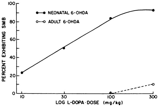
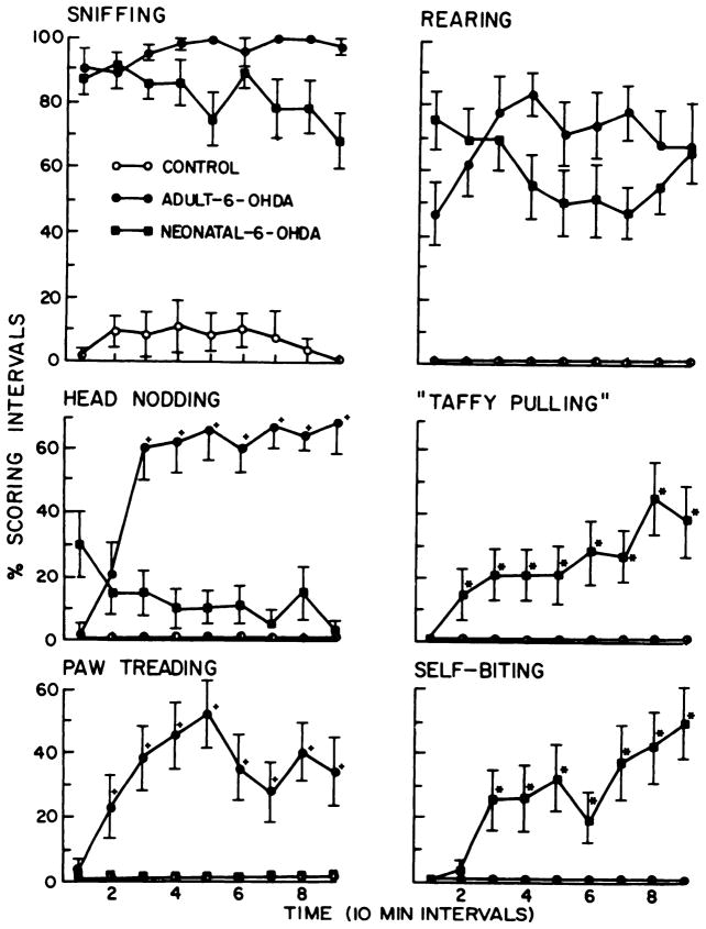
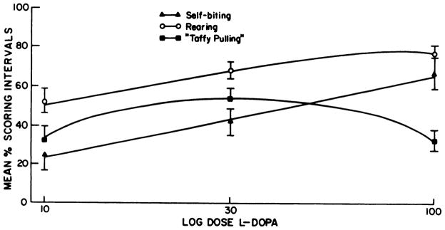
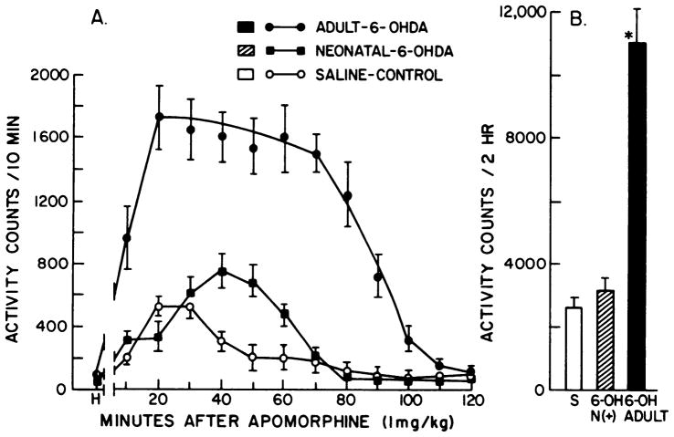
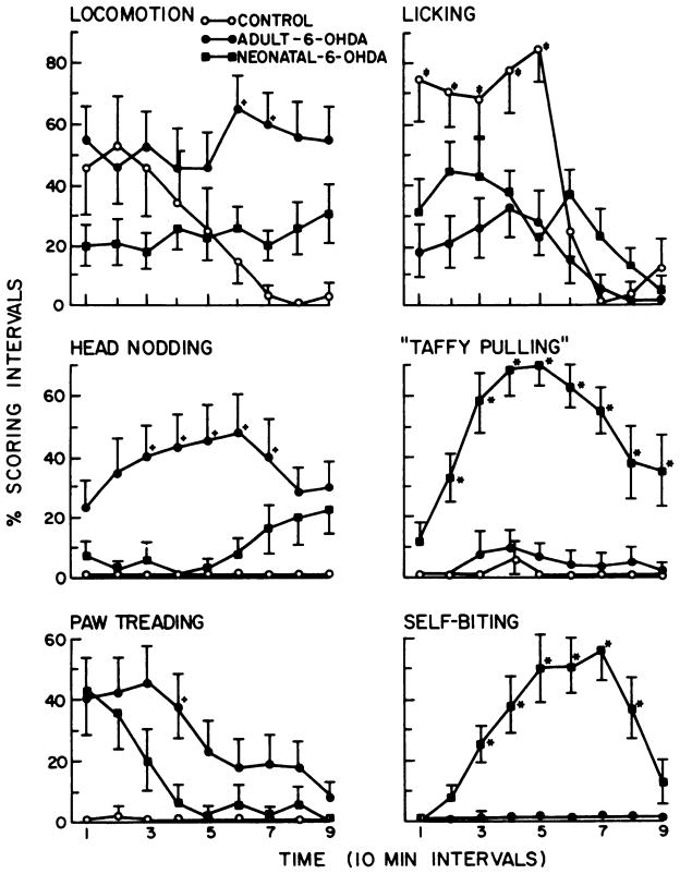
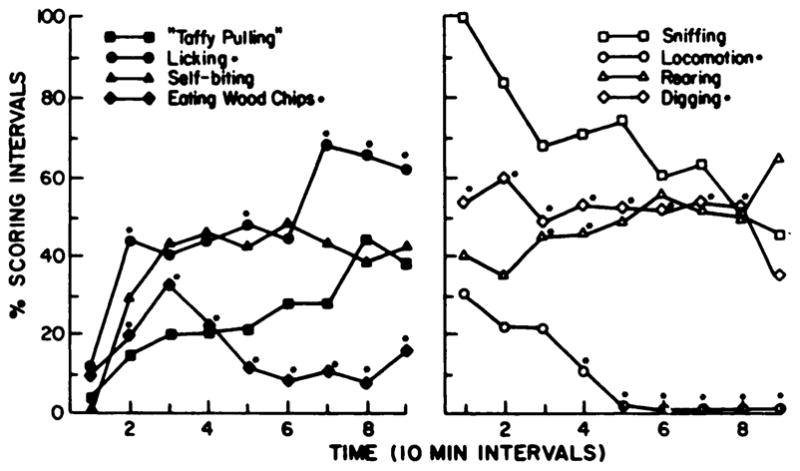
Administration of L-dopa or apomorphine to neonatal and adult 6-hydroxydopamine (6-OHDA)-treated rats resulted in different behavioral responses depending on the age at which dopaminergic fibers were destroyed. When neonatal 6-OHDA-treated rats were tested as adults, they exhibited marked stereotypies, self-biting and self-mutilation behavior (SMB) when given these dopamine agonists. Self-biting as well as the incidence of SMB in neonatal 6-OHDA-treated rats showed dose-related changes between 10 and 100 mg/kg of L-dopa. This SMB and self-biting after L-dopa was observed as early as 22 to 24 days of age. Adult 6-OHDA-treated rats did not exhibit SMB or self-biting to L-dopa (100 mg/kg) or apomorphine (10 mg/kg), but did display paw treading and head nodding--behaviors not observed in neonatal 6-OHDA-treated rats. In addition, the locomotor response to apomorphine (1 mg/kg) was significantly greater in adult 6-OHDA-treated rats than in neonatal 6-OHDA-treated rats. Brain dopamine was reduced markedly in striatum, nucleus accumbens and olfactory tubercles in both 6-OHDA treatment groups with the reduction being slightly greater in rats treated with 6-OHDA neonatally. Serotonin content was elevated in striatum of rats treated neonatally with 6-OHDA, but not in adult 6-OHDA-treated rats. SMB and behaviors observed after L-dopa in rats treated neonatally with 6-OHDA were not apparent after L-dopa in rats with brain serotonin or norepinephrine reduced. Rats with brain dopaminergic fibers destroyed neonatally exhibited self-biting and SMB after L-dopa, suggesting that neonatal reduction of this amine is responsible for the SMB and self-biting in neonatal 6-OHDA-treated rats. 5-Hydroxytryptophan administration to neonatal 6-OHDA-treated rats did not induce SMB, indicating that release of serotonin by L-dopa is not responsible for this behavior. Because inhibition of dopamine-beta-hydroxylase did not alter the SMB response to L-dopa observed in neonatal 6-OHDA-treated rats, norepinephrine synthesized from L-dopa does not appear to contribute to the response. High doses of a decarboxylase inhibitor sufficient to inhibit conversion of dopa to dopamine in brain did not reduce the incidence of SMB. Administration of haloperidol (1 mg/kg) reduced the incidence of SMB, but did not antagonize the self-biting or the taffy pulling exhibited by L-dopa. In contrast, cisflupentixol completely blocked the SMB and self-biting induced by L-dopa.(ABSTRACT TRUNCATED AT 400 WORDS)


Figures

Fig. 1
Fig. 1
Dose-response relationships of L-dopa to induce SMB in adult and neonatal 6-OHDA-treated rats. A second dose of L-dopa (100 mg/ kg) was administered to 53 of the neonatal 6-OHDA-treated rats and to 18 of the adult-treated 6-OHDA-treated rats in table 1. After this determination, 6 neonatal 6-OHDA-treated rats that were negative at 100 mg/ kg of L-dopa and 10 of the adult 6-OHDA-treated rats were given 300 mg/kg of 6-OHDA. The neonatal 6-OHDA-treated rats positive for SMB at the 100 mg/kg dose were given 30 mg/kg of L-dopa. Those SMB positive rats at 30 mg/kg of L-dopa were subsequently given 10 mg/kg of L-dopa. Percent exhibiting SMB was determined from the initial group tested (53) at the 100 mg/kg dose of L-dopa. Log L-dopa dose indicates that doses are plotted on a logarithmic scale.
Fig. 2
Fig. 2
L-dopa-induced behaviors in control and neonatal and adult 6-OHDA-treated rats. The six behaviors illustrated are all significantly different from control (P < .05). The percent scoring interval is the proportion of the four 15-sec periods in which a behavior is observed at each observation point. The time points with a (+) or an (*) indicate significant differences (P < .05) when adult and neonatal 6-OHDA-treated rats are compared. Locomotor activity (not in graph) was significantly elevated in both groups over control (P < .05), but was not different between the 6-OHDA-treated groups (P > .1). The 6-OHDA-treated groups did not dig in wood chips or eat them to any significant degree (data not graphed; see fig. 6). *, p < .05: value in the neonatal 6-OHDA treatment is significant greater than that in the adult 6-OHDA treatment group. +, p < .05: value in the adult 6-OHDA treatment is significantly greater than that in the neonatal 6-OHDA treatment group.
Fig. 3
Fig. 3
Dose-response effects of L-dopa on selected behaviors in neonatal 6-OHDA-treated rats. Behaviors were assessed as described under “Methods” at 10,30 and 100 mg/kg of L-dopa. Animals are the same as those used to make the dose-response curve for SMB in neonatal 6-OHDA-treated rats (fig. 1). Log dose L-dopa refers to doses of L-dopa on a logarithmic plot.
Fig. 4
Fig. 4
Effects of adult and neonatal 6-OHDA treatment on apomorphine-induced activity. Rats were allowed 1 hr habituation to the activity cages before receiving 1 mg/kg of apomorphine HCl s.c. Panel A shows consecutive 10-min period activity scores through 120 min. Panel B refers to the total activity counts collected for the 120 min session. Activity counts per 120 min for groups given only saline were as follows: control, 646 ± 70; adult 6-OHDA, 664 ± 170; neonatal 6-OHDA, 560 ± 165. Treatments are those described under “Methods.” H, counts accumulated during the last 10 min of habituation. S refers to saline controls that received apomorphine. 6-OH N(+) refers to neonatal 6-OHDA-treated rats positive for SMB and 6-OH adult refers to adult 6-OHDA-treated rats that received apomorphine. *p < .001 when compared to saline or to neonatal 6-OHDA-treated rats.
Fig. 5
Fig. 5
Apomorphine-induced behaviors in control and neonatal and adult 6-OHDA-treated rats. The percent scoring interval is the proportion of the four 15-sec periods in which a behavior is observed at each observation point. At some point in each graph, behavior in controls given 1 mg/kg of apormorphine is significantly different from the other treatments. The time points with a (+), an (*), or a (±) indicate significant differences (P < .05) when adult and neonatal 6-OHDA-treated groups are compared (see below). Rearing activity was not significantly different between the 6-OHDA treatment groups (P > .1), but was significantly elevated (P < .05) in both groups compared to saline treatments (data not shown). Sniffing resembled the response pattern for licking [data not shown; i.e., 6-OHDA treatment responses were not significantly different, but were significantly increased (P < .05) compared to saline]. Duration for both was greater than for control. *, P < .05: value in the neonatal 6-OHDA-treated rats is significantly greater than the corresponding one in the adult 6-OHDA-treatment group. +, P < .05: value in the adult 6-OHDA-treated rats is significantly greater than the corresponding one in the neonatal 6-OHDA treatment group. ±, P < .05: value in saline-control group is significantly greater than the corresponding values for both 6-OHDA treatment groups.
Fig. 6
Fig. 6
Effects of neonatal 6-OHDA-treatment causing preferential reduction of brain dopamine on behavioral responses to L-dopa. Behaviors illustrated are in rats treated with desipramine (20 mg/kg) 1 hr before 6-OHDA (100 μg intracistemally) to reduce only brain dopamine and not norepinephrine at 5 days of age (see table 5). Behaviors were scored as described under “Methods.” Over time, behaviors induced by L-dopa (100 mg/kg) in rats with brain dopamine preferentially reduced were compared statistically to behavioral responses observed in 6-OHDA-treated neonates, controls and adult 6-OHDA-treated rats. Behaviors illustrated are significantly different from control. Notations on the graph refer to differences with behaviors observed in neonatal 6-OHDA-treated rats with both catecholamines reduced (see fig. 2 for comparison with some of the behaviors). *p < .05 when compared to neonatal 6-OHDA-treated rats.

References

    1. Breese GR, COOPER BR. Behavioral and biochemical interactions of 5,7-dihydroxytryptamine with various drugs when administered intracisternally to adult and developing rats. Brain Res. 1975;98:517–527. - PMC - PubMed
    1. Breese GR, Traylor TD. Effects of 6-hydroxydopamine on brain norepinephrine and dopamine: Evidence for selective degeneration of catecholamine neurons. J Pharmacol Exp Ther. 1970;174:413–420. - PMC - PubMed
    1. Breese GR, Traylor TD. Developmental characteristics of brain catecholamines and tyrosine hydroxylase in the rat: Effects of 6-hydroxydopamine. Br J Pharmacol. 1972;44:210–222. - PMC - PubMed
    1. Breese GR, Cooper BR, Mueller RA. Evidence for an involvement of 5-hydroxytryptamine in the actions of amphetamine. Br J Pharmacol. 1974;52:307–319. - PMC - PubMed
    1. Breese GR, Cooper BR, Smith RD. Biochemical and behavioral alterations following 6-hydroxydopamine administration into brain. In: Usdin E, Snyder S, editors. Frontiers in Catecholamine Research. Pergamon Press; New York: 1973a. pp. 701–706.

Publication types

MeSH terms

LinkOut - more resources