

A synaptic basis for T-lymphocyte activation
- PMID: 6151375
- PMCID: PMC2551763
- DOI: 10.1016/s0769-2625(84)81105-8
A synaptic basis for T-lymphocyte activation
Abstract
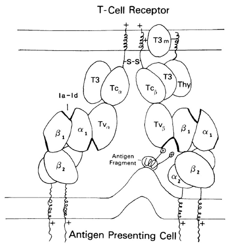
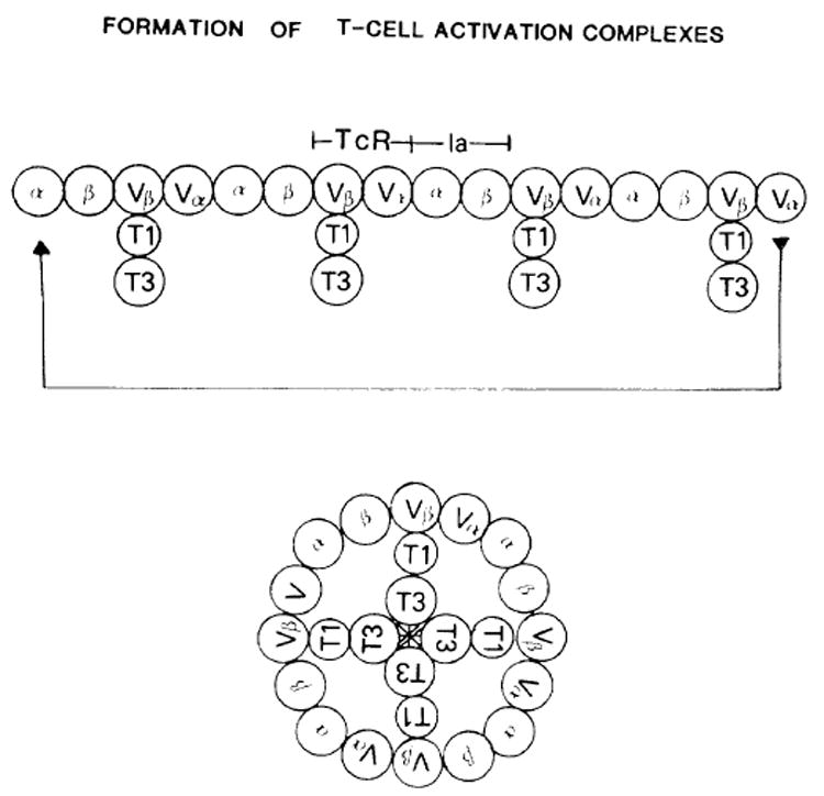
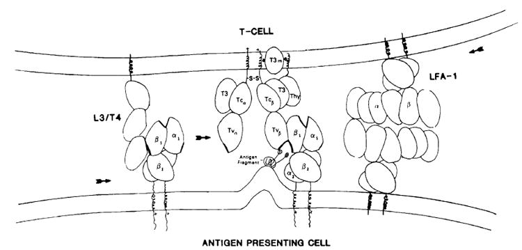
T lymphocytes respond to foreign antigen by forming specialized junctions with antigen-presenting cells (APC) or target cells. A hypothesis is presented, illustrating the similarity between the T-cell recognition-activation process and the cell communication processes found in other organ systems, especially the nervous system. Based on data showing that a major neuronal protein, Thy-1, is also a mitogenic site on T cells, and based on predictions for the structures of the T-cell receptor (TcR) and Ia proteins, an activation model is presented as follows. 1) The T-cell receptor initiates cell-cell contact with the APC by interacting with Ia and antigen, forming an antigen-binding site. 2) Sets of adhesion molecules then bind, focusing the interacting proteins to the junctional site. One binding protein, L3/T4, binds Ia and concentrates the Ia molecules to the contact site. 3) The two-chain TcR then links together the TcR-Ia-antigen complexes, forming a linear chain of receptors which will self-associate once reaching a critical length, forming a cluster. This cluster juxtaposes associated channel subunits, the T3 membrane molecules, creating an ion channel, stimulating the T cell. 4) The MHC molecule is structurally a part of this activation complex, and therefore also forms a cluster on the APC surface, possibly activating the presenting cell. 5) Secretory products are then released into the synaptic site allowing for efficient and directed cell-cell communication. Cytolytic class-I-restricted cells use a similar pathway to focus the effect of cytolytic proteins. This analogy views neuronal communication and lymphoid recognition as evolutionary descendents of a primordial lymphocytic type of cell interaction.
Figures
References
-
- McClain LD, Tomana M, Acton RT. Purification and characterization of mouse brain Thy1.2 differentiation alloantigen. Brain Res. 1978;159:161–171. - PubMed
-
- Norcross MA, Smith RT. Regulation of T-cell mitogen activity of anti-lymphocyte serum by a B helper cell. J Immunol. 1979;122:1620–1628. - PubMed
-
- Maino VC, Norcross MA, Perkins MS, Smith RT. Mechanisms of Thy-1 mediated T-cell activation: roles of Fc receptors, T200, and H-2 glycoproteins in accessory cell function. J Immunol. 1981;126:1829–1836. - PubMed
-
- Konaka Y, Norcross MA, Maino VC, Smith RT. Anti-Thy-1 mediated T-cell activation. Role of soluble factors and expression of growth factor receptors on T cells. Europ J Immunol. 1981;11:445–450. - PubMed
-
- Shimizu S, Smith RT, Norcross MA, Maino VC. Mechanisms controlling TCGF production by cloned sublines of EL-4 azgr in response to stimulation by anti-Thy-1 antibody. J Immunol. 1982;128:296–301. - PubMed
Publication types
MeSH terms
Substances
Grants and funding
LinkOut - more resources
Full Text Sources
Research Materials
Miscellaneous