. 1983 Jun;303(5916):446-8.
doi: 10.1038/303446a0.

Polyoma virus 'hexamer' tubes consist of paired pentamers

Polyoma virus 'hexamer' tubes consist of paired pentamers

T S Baker et al. Nature. 1983 Jun.

Abstract

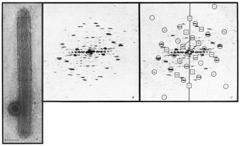
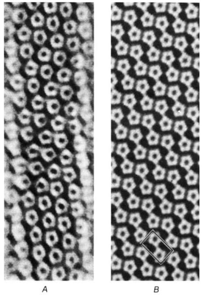
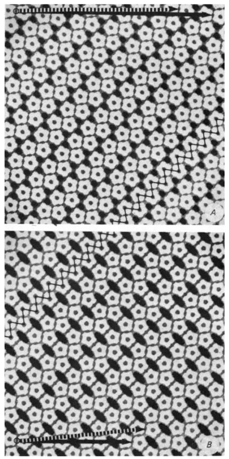
The discovery that the 72 capsomeres of the icosahedrally symmetric polyoma virus capsid are all pentamers shows that the expected quasi-equivalent bonding specificity is not conserved in the assembly of this virus coat protein. Tubular particles produced by polyoma and other papovaviruses seem to be polymorphic aggregates of capsomeres that may arise through variation in switching of the bonding specificity. Electron micrographs of wide and narrow classes of tubes were analysed by Kiselev and Klug using optical diffraction and optical filtering methods. The wide type were called 'hexamer' tubes because they consist of approximately hexagonally arrayed capsomeres that were assumed to be hexamers, in accord with the quasi-equivalence theory of icosahedral virus particle construction. The narrow type were called 'pentamer' tubes because the capsomeres are arrayed in a particular 'pentagonal tesselation' which arises from the pairing of pentamers across 2-fold axes of the surface lattice. Our reexamination of negatively-stained polyoma virus tubes by digital image processing of low-irradiation electron micrographs shows that all tubes are assemblies of paired pentameric capsomeres. We report here that the packing arrangement of the pentamers in the hexamer tubes is simply related to the pentagonal tessellation respresenting the packing in the narrow pentamer tubes. In all the tube structures examined, at least one pairwise contact between neighbouring pentamers closely resembles the contact between the pentavalent and hexavalent capsomeres in the icosahedral capsid.


Figures

Fig. 1
Fig. 1
Indexing of the diffraction pattern from a polyoma virus hexamer tube image shows that the true unit cell is twice as large as the simple hexagonal cell previously described The electron micrograph (A) is representative of the most frequent class of hexamer tube. The tube, capped at both ends, appears uniformly flattened except where it is supported by the adjacent icosahedral capsid. Superposition of the front and back lattices gives rise to a characteristic criss-cross pattern of near axial rows of capsomeres. Scale bar, 500 Å. The diffraction pattern computed from the area boxed in A is shown in B and again in C with indexing of the spots arising from the top layer (away from the grid) of the flattened tube. Circles indicate spots from the simple hexagonal unit cell, which would contain only one capsomere, and the squares mark additional spots from the repeating unit of double this size. Spots from the bottom side (not marked) lie on a lattice related to that from the top by approximate mirror symmetry about the meridional axis (dashed line). The reciprocal lattice unit cell for the top layer is marked at the centre of the pattern.
Fig.2
Fig.2
The filtered image of the front layer of a flattened hexamer tube corresponds to a model surface lattice built of pairs of pentamers arrayed with p2 symmetry. Image A was computed from the diffraction pattern of the portion of the tube boxed in Fig. 1A. The pattern was filtered to include the spots indexed in Fig. 1C and the equator (with the equator and the meridional pair of spots given half weighting). The planar model (B) was built in the computer from regular pen tamers arranged in a p2 lattice at the coordinates measured for the capsomeres in the unit cell of the filtered image. One choice of unit cell is boxed.
Fig.3
Fig.3
Comparison of the pentamer packing in hexamer (A) and pentamer (B) tube surface lattices illustrates correspondences in bonding contacts. The ‘zig’ and ‘zag’ lines (oriented respectively nearly vertically and horizontally along the diagonally directed ribbon) indicate the two classes of dimer ‘bonds’ between pentamers that are conserved in the hexamer and pentamer tube lattices. These planar models were constructed with equivalent connections for the differently oriented edge-to-edge bonds between the pentagonal units, and the packing width of the zigzag ribbon in A was made the same as in B. In the cylindrically curved tube surface, the differently oriented edge-to-edge contacts are non-equivalently bent. Furthermore, the relative separations of capsomeres in the different bonding directions measured from electron micrographs are slightly distorted compared to these idealized models. Circumferential vectors corresponding to different tube structures are marked by arrows between equivalent lattice points. Models of the tubes can be constructed from these plane lattices by cutting out rectangles based on the circumferential vectors and connecting the vertical edges to form cylinders. Two of the frequently observed size of hexamer tubes are indicated by the arrows at the top of A. the dashed one representing the circumference of the tube in Fig. 1A. Hexamer tubes of larger and smaller diameter have circumferential vectors in approximately the same direction as the marked arrows. The circumferential vectors of the zero- and one-start pentamer tubes (which are the only identified narrow tubes of this type) are marked by the solid and dashed arrows, respectively, at the bottom of B.

References

    1. Rayment I, Baker TS, Caspar DLD, Murakami WT. Nature. 1982;295:110–115. - PMC - PubMed
    1. Caspar DLD, Klug A. Cold Spring Harb. Symp. quant. Biol. 1962;27:1–24. - PubMed
    1. Finch JT, Klug A. I. molec. Biol. 1965;13:1–12. - PubMed
    1. Kiselev NA, Klug A. I. molec. Biol. 1969;40:155–171. - PubMed
    1. Baker TS, Amos LA. I. molec. Biol. 1978;123:89–106. - PubMed

Publication types