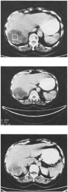
Accuracy of computerized tomography in determining hepatic tumor size in patients receiving liver transplantation or resection
- PMID: 6327930
- PMCID: PMC3154766
- DOI: 10.1200/JCO.1984.2.6.637
Accuracy of computerized tomography in determining hepatic tumor size in patients receiving liver transplantation or resection
Abstract
Computerized tomography (CT) of liver is used in oncologic practice for staging tumors, evaluating response to treatment, and screening patients for hepatic resection. Because of the impact of CT liver scan on major treatment decisions, it is important to assess its accuracy. Patients undergoing liver transplantation or resection provide a unique opportunity to test the accuracy of hepatic-imaging techniques by comparison of findings of preoperative CT scan with those at gross pathologic examination of resected specimens. Forty-one patients who had partial hepatic resection (34 patients) or liver transplantation (eight patients) for malignant (30 patients) or benign (11 patients) tumors were evaluable. Eight (47%) of 17 patients with primary malignant liver tumors, four (31%) of 13 patients with metastatic liver tumors, and two (20%) of 10 patients with benign liver tumors had tumor nodules in resected specimens that were not apparent on preoperative CT studies. These nodules varied in size from 0.1 to 1.6 cm. While 11 of 14 of these nodules were less than 1.0 cm, three of 14 were greater than 1.0 cm. These results suggest that conventional CT alone may be insufficient to accurately determine the presence or absence of liver metastases, extent of liver involvement, or response of hepatic metastases to treatment.
Figures
References
-
- Moertel CG, Hanley JA. The effect of measuring error on the results of therapeutic trials in advanced cancer. Cancer. 1976;38:388–394. - PubMed
-
- Kaplan BH. Measuring the Effect of Anti-Cancer Treatment. Wilmington: Stuart Pharmaceuticals; 1981.
-
- Lavin PT, Flowerdew G. Studies in variation associated with the measurement of solid tumors. Cancer. 1980;46:1286–1290. - PubMed
-
- Ettinger DS, Leichner PK, Siegelman S, et al. Computed tomography (CT) associated volumetric analysis of primary liver tumors as a measure of response to therapy. Proc Am Soc Clin Oncol. 1983;2:115. Abstr. - PubMed
-
- Bernadino ME, Lewis E. Imaging hepatic neoplasms. Cancer. 1982;50:2666–2671. - PubMed
MeSH terms
Grants and funding
LinkOut - more resources
Full Text Sources
Medical


