. 1981 Jan 25;256(2):575-8.

Mechanism of action of troponin . tropomyosin. Inhibition of actomyosin ATPase activity without inhibition of myosin binding to actin

Mechanism of action of troponin . tropomyosin. Inhibition of actomyosin ATPase activity without inhibition of myosin binding to actin

J M Chalovich et al. J Biol Chem. .

Abstract

The regulation of vertebrate skeletal muscle contraction by the troponin . tropomyosin complex is generally thought to be the result of tropomyosin physically blocking the myosin binding site of actin in the absence of Ca2+. This mechanism was tested during steady state ATP hydrolysis by comparing the degree of association of myosin subfragment 1 (S-1) with the actin . troponin . tropomyosin complex in the absence and presence of Ca2+. Binding in the presence of ATP was determined by stopped flow absorbance measurements at 25 degrees C. Although the steady state ATPase rate was reduced 96% in the absence of Ca2+, the association constant of S-1 with regulated actin was virtually the same in the absence of Ca2+ (1.3 X 10(4) M-1) as in the presence of Ca2+ (2.3 X 10(4) M-1). The association constant of S-1 to regulated actin in the presence of Ca2+ was similar to the association constant of S-1 to unregulated actin. These results suggest that the troponin . tropomyosin complex does not inhibit the actin-activated ATPase activity by preventing the binding of S-1 . ATP or S-1 . ADP . Pi to actin; rather, it may act by blocking the release of Pi from the acto-S-1 . ADP . Pi complex.


Figures

Fig. 1
Fig. 1. Final absorbance of the complex formed upon binding of S-1 to actin or regulated actin as a function of S-1 concentration
The final absorbance was measured after completion of ATP hydrolysis. Conditions: 78 μm actin, 17 μm native tropomyosin (where applicable), 1.0 mm ATP, 3.0 mm MgCl2, 1 mm EGTA, 10 mm imidazole, pH 7.0, 25°C. •, ▪, stopped flow measurement; ○, standard spectrophotometer measurement.
Fig. 2
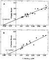
Fig. 2. Double reciprocal plots of the fraction of S-1 bound, in the presence of ATP, as a function of free actin concentration
The conditions are the same as in Fig. 1 with 20 μm S-1, except where noted. A, binding in the presence of calcium. EGTA was replaced with 0.5 mm CaCl. Binding to regulated (closed symbols) and unregulated actin (open symbols) are shown. B, binding in the absence of calcium. S-1 concentration was varied from 10 μm (▴) to 40 μm (▵). In both A and B, different symbols represent different protein preparations. The solid lines are least squares fits to the data (see “Materials and Methods”).
Fig. 3
Fig. 3. Time course of the fraction of S-1 bound to actin or regulated actin during ATP hydrolysis
Conditions are the same as in Fig. 1 with 20 μm S-1 and free actin concentrations of 90 μm (regulated + Ca2+), 100 μm (unregulated), and 130 μm (regulated + EGTA).
Fig. 4
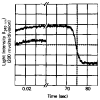
Fig. 4. Time course of the transmitted light intensity after mixing regulated actin with either S-1 (upper curve) or buffer (lower curve) in the presence of ATP and EGTA
The lower curve has been moved down about 1 V for comparison. Conditions are the same as in Fig. 1 with the following final protein concentrations: S-1, 20 μm; actin, 60 μm; native tropomyosin, 13 μm. 35% of the S-1 was bound to actin during steady state ATP hydrolysis in this experiment.

References

    1. Chalovich JM, Chock PB, Eisenberg E. Fed Proc. 1980;39:1106. (abstr.)
    1. Huxley AF. Prog Biophys Biophys Chem. 1957;7:255–318. - PubMed
    1. Huxley AF. J Physiol Land. 1974;243:1–43. - PMC - PubMed
    1. Huxley HE. Science. 1969;164:1356–1366. - PubMed
    1. Perry SV. Biochem Soc Trans. 1979;7:593–617. - PubMed